Antitragicus
| Antitragicus | |
|---|---|
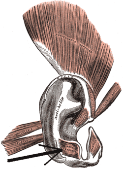 The muscles of the ear (antitragicus visible at bottom center). | |
| Details | |
| Origin | Outer part of the antitragus |
| Insertion | Cauda helicis and antihelix |
| Artery | Auricular branch of superficial temporal and auricular branches of posterior auricular artery |
| Nerve | Facial nerve |
| Actions | Modifies the auricular shape |
| Identifiers | |
| Latin | Musculus antitragicus |
| TA | A15.3.01.042 |
| FMA | 48980 |
The Antitragicus is an intrinsic muscle of the outer ear.
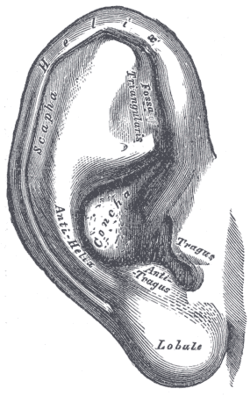
In human anatomy, the antitragicus arises from the outer part of the antitragus, and is inserted into the cauda helicis (or tail of the helix) and antihelix.[1][2]
The function of the muscle is to adjusts the shape of the ear by pulling the antitragus and cauda helicis towards each other. While the muscle modifies the auricular shape only minimally in the majority of individuals, this action could increase the opening into the external acoustic meatus in some.[1]
The helicis minor is developmentally derived from the second pharyngeal arch.[1]
Additional images
|
See also
References
This article incorporates text in the public domain from the 20th edition of Gray's Anatomy (1918)
- Bennett S, Dagash H, McArthur P (2005). "The role of the antitragicus muscle in plical folding of the pinna.". Plast Reconstr Surg. 115 (5): 1266–8. doi:10.1097/01.PRS.0000156773.69631.52. PMID 15809584.
- 1 2 3 "Antitragus". AnatomyExpert. Archived from the original on 10 May 2013. Retrieved 9 March 2013.
- ↑ "Henry Gray (1825–1861). Anatomy of the Human Body. 1918.". Bartleby.com. Retrieved 9 March 2013.
External links
This article is issued from Wikipedia - version of the 10/15/2016. The text is available under the Creative Commons Attribution/Share Alike but additional terms may apply for the media files.