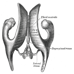
Body of lateral ventricle
| Body of lateral ventricle | |
|---|---|
 Drawing of a cast of the ventricular cavities, viewed from above. | |
 Drawing of a cast of the ventricular cavities, viewed from the side. | |
| Details | |
| Identifiers | |
| Latin | Pars centralis ventriculi lateralis |
| NeuroNames | hier-203 |
| NeuroLex ID | Body of lateral ventricle |
| TA | A14.1.09.274 |
| FMA | 83703 |
The body of lateral ventricle (cella) extends from the interventricular foramen to the splenium of the corpus callosum.
It is an irregularly curved cavity, triangular on transverse section, with a roof, a floor, and a medial wall.
The roof is formed by the under surface of the corpus callosum; the floor by the following parts, enumerated in their order of position, from before backward: the caudate nucleus of the corpus striatum, the stria terminalis and the terminal vein, the lateral portion of the upper surface of the thalamus, the choroid plexus, and the lateral part of the fornix; the medial wall is the posterior part of the septum pellucidum, which separates it from the opposite ventricle.
Additional images
-

Dissected human brain seen from the right.
References
This article incorporates text in the public domain from the 20th edition of Gray's Anatomy (1918)
External links
- Atlas image: n1a4p2 at the University of Michigan Health System