Bright-field microscopy

Bright-field microscopy is the simplest of all the optical microscopy illumination techniques. Sample illumination is transmitted (i.e., illuminated from below and observed from above) white light and contrast in the sample is caused by absorbance of some of the transmitted light in dense areas of the sample. Bright-field microscopy is the simplest of a range of techniques used for illumination of samples in light microscopes and its simplicity makes it a popular technique. The typical appearance of a bright-field microscopy image is a dark sample on a bright background, hence the name.
Light path
The light path of a bright-field microscope is extremely simple, no additional components are required beyond the normal light microscope setup. The light path therefore consists of:
- a transillumination light source, commonly a halogen lamp in the microscope stand;
- a condenser lens which focuses light from the light source onto the sample; and
- objective lens which collects light from the sample and magnifies the image.
- oculars and/or a camera to view the sample image
Bright field microscopy may use critical or Köhler illumination to illuminate the sample.
Performance
Bright-field microscopy typically has low contrast with most biological samples as few absorb light to a great extent. Staining is often required to increase contrast, which prevents use on live cells in many situations. Bright field illumination is useful for samples which have an intrinsic colour, for example chloroplasts in plant cells.
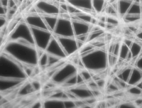
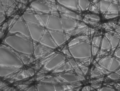
- Comparison of transillumination techniques used to generate contrast in a sample of tissue paper. 1.559 μm/pixel.
-

Bright field illumination, sample contrast comes from absorbance of light in the sample.
-

Cross-polarized light illumination, sample contrast comes from the rotation of polarized light through the sample.
-
Dark field illumination, sample contrast comes from light scattered by the sample.
-
Phase contrast illumination, sample contrast comes from interference of different path lengths of light through the sample.
Bright-field microscopy is a standard light microscopy technique, and therefore magnification is limited by the resolving power possible with the wavelength of visible light.
Advantages
- Simplicity of setup with only basic equipment required.
Limitations
- Very low contrast of most biological samples.
- Low apparent optical resolution due to the blur of out of focus material.
- Samples that are naturally colorless and transparent cannot be seen well, e.g. many types of mammalian cells. These samples often have to be stained before viewing. Samples that do have their own colour can be seen without preparation, e.g. the observation of cytoplasmic streaming in Chara cells.
Enhancements
- Reducing or increasing the amount of the light source via the iris diaphragm.
- Use of an oil immersion objective lens and a special immersion oil placed on a glass cover over the specimen. Immersion oil has the same refraction as glass and improves the resolution of the observed specimen.
- Use of sample staining methods for use in microbiology, such as simple stains (Methylene blue, Safranin, Crystal violet) and differential stains (Negative stains, flagellar stains, endospore stains).
- Use of a colored (usually blue) or polarizing filter on the light source to highlight features not visible under white light. The use of filters is especially useful with mineral samples.
References
- Advanced Light Microscopy vol. 1 Principles and Basic Properties by Maksymilian Pluta, Elsevier (1988)
- Advanced Light Microscopy vol. 2 Specialised Methods by Maksymilian Pluta, Elsevier (1989)
- Introduction to Light Microscopy by S. Bradbury, B. Bracegirdle, BIOS Scientific Publishers (1998)
- Microbiology: Principles and Explorations by Jacquelyn G. Black, John Wiley & Sons, Inc. (2005)
- Microscopy and Imaging Literature
Optical microscopy | ||
|---|---|---|
| Illumination and contrast methods |  | |
| Fluorescence methods | ||
| Sub-diffraction limit techniques | ||
External links
| Library resources about Bright-field microscopy |