Calcaneal fracture
| Calcaneal fracture | |
|---|---|
| lover's fracture, Don Juan fracture | |
|
X-ray of a fractured calcaneus. | |
| Classification and external resources | |
| Specialty | orthopedics, emergency medicine |
| ICD-10 | S92.0 |
| ICD-9-CM | 825.0-825.2 |
| eMedicine | radio/123 |
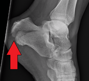
Calcaneal fracture is a fracture of the calcaneus. It is usually caused by a fall from height when one lands on their feet. These fractures represent approximately 2% of all fractures but 60% of tarsal bone fractures.[1] The name lover's fracture is derived from the fact that a lover may jump from great heights while trying to escape from the lover's spouse.[2]
Signs and symptoms
The most common symptom is pain over the heel area, especially when the heel is palpated or squeezed. Patients usually have a history of recent trauma to the area or fall from a height. Other symptoms include: inability to bear weight over the involved foot, limited mobility of the foot, and limping. Upon inspection, the examiner may notice swelling, redness, and hematomas. A hematoma extending to the sole of the foot is called "Mondor Sign", and is pathognomonic for calcaneal fracture.[3][4] The heel may also become widened with associated edema due to displacement of lateral calcaneal border. Involvement of soft tissue (tendons, skin, etc.,) should be evaluated because soft tissue injury has been associated to serious complications (see below).[5][6]
Cause
Calcaneal fractures are often attributed to shearing stress adjoined with compressive forces combined with a rotary direction (Soeur, 1975[7]). These forces are typically linked to injuries in which an individual falls from a height, involvement in an automobile accident, or muscular stress where the resulting forces can lead to the trauma of fracture. Overlooked aspects of what can lead to a calcaneal fracture are the roles of osteoporosis and diabetes.
Unfortunately, prevention towards falls and automobile accidents is limited and applies to unique circumstances that should be avoided. The aspect of muscular stress fractures can be prevented through strength training and stretching such as weight bearing exercises. In addition, footwear can create an impact on the forces that may cause a calcaneal fracture and can prevent them as well. A study conducted by Salzler(2012[8]), exposed that the increasing trend of minimalist footwear or running barefoot can lead to a variety of stress fractures including that of calcaneal fractures.
Osteoporosis
As the population ages, their bone mineral density decreases. The process of bone loss in the condition of osteoporosis can be prevented through an adequate intake of vitamin C and vitamin D coupled with exercise and by being a non-smoker. A study by Cheng et al. in 1997,[9] showed that greater bone density indicated less risk for fractures in the calcaneus.
Diabetes
In 1991, Kathol[10] conducted a study which helped show the correlation between calcaneal insufficiency avulsion fractures (A fracture in which the Achilles tendon will remove a portion of the bone when it rescinds) and diabetes mellitus. The diabetic population that suffers from the disease is more susceptible to the risks of fracture and potentially healing complications and infection that may lead to limb amputation. Diabetes can be regulated and prevented through diet and exercise.
Diagnosis

Conventional radiography is usually the initial assessment tool when calcaneal fracture is suspected. Recommended x-ray views are (a) axial, (b) anteroposterior, (c) oblique views and (d) views with dorsiflexion and internal rotation of the foot. However, conventional radiography is limited for visualization of calcaneal anatomy, especially at the subtalar joint. CT Scan is currently the imaging study of choice for evaluating calcaneal injury and has substituted conventional radiography in the classification of calcaneal fractures.[11] Axial and coronal views are taken for proper visualization of the calcaneus, subtalar, calcaneocuboid and talonavicular joints.
Classification

The calcaneus, also known as the heel bone, is the largest of the tarsal bones and articulates with the cuboid bone anteriorly and the talus bone superiorly. It is responsible for transmitting the majority of the body's weight from the talus bone to the ground.
Calcaneal fractures are categorized into two types: Intra- and Extrarticular fractures on the basis of subtalar joint involvement. Intrarticular fractures are more common and involve the posterior talar articular facet of the calcaneus. The Sanders classification groups these fractures into four types, based on the location of the fracture at the posterior articular surface. Extrarticular fractures are less common, and located anywhere outside the subtalar joint.[11] Extrarticular fractures are categorized depending on whether the involvement of the calcaneus is anterior (Type A), Middle (Type B) or Posterior (Type C).
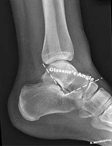
The Angle of Gissane, or "Critical Angle", is the angle formed by the downward and upward slopes of the calcaneal superior surface. On a lateral radiograph, an angle of Gissane of > 130° suggests fracture of the posterior subtalar joint surface. Bohler's angle, or the "Tuber Angle", is another normal anatomic landmark seen in lateral radiographs. It is formed by the intersection of 1) a line from the highest point of the posterior articular facet to the highest point of the posterior tuberosity, and 2) a line from the former to the highest point on the anterior articular facet. An angle < 20° suggests a depression of posterior facet and possible calcaneal fracture.
The Sanders classification system is the most commonly used system for categorizing intrarticular fractures. There are 4 types:
- Type I fractures are non-displaced fractures (displacement < 2 mm).
- Type II fractures consist of a single intrarticular fracture that divides the calcaneus into 2 pieces.
- Type IIA: fracture occurs on lateral aspect of calcaneus.
- Type IIB: fracture occurs on central aspect of calcaneus.
- Type IIC: fracture occurs on medial aspect of calcaneus.
- Type III fractures consist of two intrarticular fractures that divide the calcaneus into 3 articular pieces.
- Type IIIAB: two fracture lines are present, one lateral and one central.
- Type IIIAC: two fracture lines are present, one lateral and one medial.
- Type IIIBC: two fracture lines are present, one central and one medial.
- Type IV fractures consist of fractures with more than three intrarticular fractures.
Extrarticular fractures include all fractures that do not involve the posterior facet of the subtalar joint.
- Type A involve the anterior calcaneus
- Type B involve the middle calcaneus. This includes the sustentaculum tali, trochlear process and lateral process.
- Type C involve the posterior calcaneus, the posterior tuberosity and medial tubercle included.
Treatment
Non-surgical treatment is indicated for extrarticular fractures and Sanders Type I intrarticular fractures, provided that the calcaneal weight-bearing surface and foot function are not compromised. Physicians may choose to perform closed reduction with or without fixation (casting), or fixation alone (without reduction), depending on the individual case. Recommendations include no weight-bearing for a few weeks followed by range-of-motion exercises and progressive weight bearing for a period of 2–3 months.
Displaced intrarticular fractures require surgical intervention within 3 weeks of fracture, before bone consolidation has occurred. Conservative surgery consists of closed reduction with percutaneous fixation. This technique is associated with less wound complications, better soft tissue healing (because of less soft tissue manipulation) and decreased intraoperative time. However, this procedure has increased risk of inadequate calcaneal bone fixation, compared to open procedures.[12] Currently, open reduction with internal fixation (ORIF) is usually the preferred surgical approach when dealing with displaced intrarticular fractures. Newer, more innovative surgical techniques and equipment have decreased the incidence of intra- and post-operative complications.
Rehabilitation
Rehabilitation for a calcaneal fractures is dependent on whether surgery was required or not. Both types of rehabilitation require three phases in which only the first phase is different.


Exercises that can be used for the range of motion phase can include eversion and inversion of the ankle, flexion and extension of the ankle, and a combination of the two motions to create a circular foot motion. Exercises that allow slight to full body weight to be used in the final phases include stepping forward then back, side-stepping, and leg stand.
Phases
The first phase of the rehabilitation after surgery includes keeping the foot elevated and iced for the first 2 days after the operation. After those 2 days, using crutches or a wheelchair in which there is no weight applied to the affected foot is recommended to getting around. If no operation was performed, the foot should be submitted to frequent range of motion exercises.[13] The second phase occurs 6 weeks after and consists of keeping the foot elevated and iced while resting and performing exercises in which only slight weight is applied to the affected area for the next two weeks, others recommend six weeks of this phase.[14] In this phase range of motion exercises should be implemented if surgery was needed for the fracture. The third and final phase of rehabilitation of calcaneal fractures is to allow the full body weight to be used and use crutches or a cane if needed, between 13 weeks to a year the patient is allowed to resume normal activities.[12]
Complications
Evaluating for soft-tissue involvement is the most important aspect of the clinical examination because of its association to patient outcome.[6][7] Skin blisters may become infected if medical attention is delayed, which can lead to necrotizing fasciitis or osteomyelitis, causing irreparable muscle/bone damage. Ligament and tendon involvement should also be explored. Achilles tendon injury can be seen with posterior (Type C) fractures. Worth mentioning is the fact that, since calcaneal fractures are related to height falls, other concomitant injuries should be sought. Vertebral compression fractures occur in approximately 10% of these patients.[1] A trauma-focused clinical approach should be implemented; tibial, knee, femur, hip, and head injuries should be ruled out by means of history and physical exam.
References
- 1 2 Stoller, D. W.; Tirman, P. F. J.; Bredella, M. (2004). "Ankle and foot, osseous fractures, calcaneal fractures". Diagnostic imaging: orthopaedics. Salt Lake City: Amirsys. pp. 70–4.
- ↑ Lee, Patrick; Hunter, Tim B.; Taljanovic, Mihra (2004). "Musculoskeletal Colloquialisms: How Did We Come Up with These Names?". Radiographics. 24 (4): 1009–27. doi:10.1148/rg.244045015. PMID 15256625.
- ↑ Calcaneus Fractures at eMedicine
- ↑ Richman, JD; Barre, PS (1986). "The plantar ecchymosis sign in fractures of the calcaneus". Clinical Orthopaedics and Related Research (207): 122–5. doi:10.1097/00003086-198606000-00022. PMID 3720074.
- ↑ Berry, G. K.; Stevens, D. G.; Kreder, H. J.; McKee, M.; Schemitsch, E.; Stephen, D. J. G. (2004). "Open Fractures of the Calcaneus: A Review of Treatment and Outcome". Journal of Orthopaedic Trauma. 18 (4): 202–6. doi:10.1097/00005131-200404000-00002. PMID 15087962.
- 1 2 Heier, Keith A.; Infante, Anthony F.; Walling, Arthur K.; Sanders, Roy W. (2003). "Open Fractures of the Calcaneus: Soft-Tissue Injury Determines Outcome". The Journal of Bone and Joint Surgery. American Volume. 85–A (12): 2276–82. PMID 14668494.
- 1 2 Soeur, Robert; Remy, Robert (1975). "Fractures of the calcaneus with displacement of the thalamic portion". The Journal of Bone and Joint Surgery. British Volume. 57 (4): 413–21. PMID 1194308.
- ↑ Salzler, Matthew J.; Bluman, Eric M.; Noonan, Samantha; Chiodo, Christopher P.; de Asla, Richard J. (2012). "Injuries Observed in Minimalist Runners". Foot & Ankle International. 33 (4): 262–6. doi:10.3113/FAI.2012.0262. PMID 22735197.
- ↑ Cheng, Sulin; Suominen, Harri; Sakari-Rantala, Ritva; Laukkanen, Pia; Avikainen, Veikko; Heikkinen, Eino (1997). "Calcaneal Bone Mineral Density Predicts Fracture Occurrence: A Five-Year Follow-up Study in Elderly People". Journal of Bone and Mineral Research. 12 (7): 1075–82. doi:10.1359/jbmr.1997.12.7.1075. PMID 9200007.
- ↑ Kathol, M H; el-Khoury, G Y; Moore, T E; Marsh, J L (1991). "Calcaneal insufficiency avulsion fractures in patients with diabetes mellitus". Radiology. 180 (3): 725–9. doi:10.1148/radiology.180.3.1871285. PMID 1871285.
- 1 2 Badillo, Kenneth; Pacheco, Jose A.; Padua, Samuel O.; Gomez, Angel A.; Colon, Edgar; Vidal, Jorge A. (2011). "Multidetector CT Evaluation of Calcaneal Fractures". RadioGraphics. 31 (1): 81–92. doi:10.1148/rg.311105036. PMID 21257934.
- 1 2 Calcaneus Fractures~treatment at eMedicine
- ↑ Godges, Joe; Klingman, Robert. "Calcaneal Fracture and Rehabilitation" (PDF). Kaiser Permanente.
- ↑ Hatzokos, I; Karataglis, D; Papadopoulos, P; Dimitriou, C; Christodoulou, A; Pournaras, J (2006). "Treatment of intra-articular comminuted os calcis fractures". Orthopedics. 29 (1): 25–9. PMID 16429931.
Bibliography
- Brant, W.; Helms, C (2007). Fundamentals of Diagnostic Radiology (3rd ed.). Lippincott Williams & Wilkins. ISBN 0-7817-6135-2.