Common bile duct
| Common bile duct | |
|---|---|
 Diagram of the biliary tree showing the common bile duct | |
| Details | |
| Identifiers | |
| Latin | ductus choledochus |
| Acronym(s) | CBD |
| FMA | 14667 |
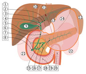
9. Gallbladder, 10–11. Right and left lobes of liver. 12. Spleen.
13. Esophagus. 14. Stomach. 15. Pancreas: 16: Accessory pancreatic duct, 17: Pancreatic duct.
18. Small intestine: 19. Duodenum, 20. Jejunum
21–22: Right and left kidneys (silhouette).
The anterior border of the liver is lifted upwards (brown arrow). Gallbladder with Longitudinal section, pancreas and duodenum with frontal one. Intrahepatic ducts and stomach in transparency.
The common bile duct, sometimes abbreviated CBD,[1] is a duct in the gastrointestinal tract of organisms that have a gall bladder. It is formed by the union of the common hepatic duct and the cystic duct (from the gall bladder). It is later joined by the pancreatic duct to form the ampulla of Vater. There, the two ducts are surrounded by the muscular sphincter of Oddi.
When the sphincter of Oddi is closed, newly synthesized bile from the liver is forced into storage in the gall bladder. When open, the stored and concentrated bile exits into the duodenum. This conduction of bile is the main function of the common bile duct. The hormone cholecystokinin, when stimulated by a fatty meal, promotes bile secretion by increased production of hepatic bile, contraction of the gall bladder, and relaxation of the Sphincter of Oddi.
Several problems can arise within the common bile duct. If clogged by a gallstone, a condition called choledocholithiasis can result.[2] In this clogged state, the duct is especially vulnerable to an infection called ascending cholangitis. Very rare deformities of the common bile duct are cystic dilations (4 cm), choledochoceles (cystic dilation of the ampula of Vater (3–8 cm), and biliary atresia.
Additional images
-
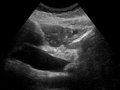
Dilatation of CBD due to Ampullary tumor.
-

The gall-bladder and bile ducts laid open.
References
- ↑ Agabegi, Steven S.; Agabegi, Elizabeth D. (23 August 2012). Step-Up to Medicine. Lippincott Williams & Wilkins. p. 136. ISBN 9781609133603.
- ↑ Humes, H. David (2001). Kelley's Essentials of Internal Medicine. Lippincott Williams & Wilkins. p. 229. ISBN 978-0781719377.
- S.E.Miederer et al.:Endoscopic transpapillary splitting of a choledochocele. Dtsch Med. Wochenschr. 1978 Feb.3:103(5):216,219. PMID 631041
External links
- Anatomy figure: 38:06-08 at Human Anatomy Online, SUNY Downstate Medical Center - "The gallbladder and extrahepatic bile ducts."
- Anatomy image:8336 at the SUNY Downstate Medical Center
- Anatomy image:7957 at the SUNY Downstate Medical Center
- liver at The Anatomy Lesson by Wesley Norman (Georgetown University) (biliarysystem)