Diffuse idiopathic skeletal hyperostosis
| Diffuse idiopathic skeletal hyperostosis | |
|---|---|
| Synonyms | Forestier's disease, senile ankylosing spondylosis, ankylosing hyperostosis |
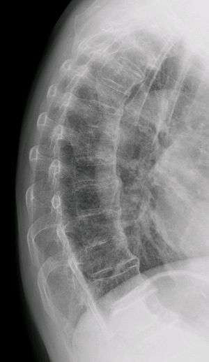 | |
| Diffuse idiopathic skeletal hyperostosis. Woman of 80 years old, also with Th11 fracture. | |
| Classification and external resources | |
| Specialty | rheumatology |
| ICD-10 | M48.1 |
| ICD-9-CM | 721.6 |
| OMIM | 106400 |
| DiseasesDB | 4932 |
| eMedicine | article/1258514 article/388973 |
| Patient UK | Diffuse idiopathic skeletal hyperostosis |
Diffuse idiopathic skeletal hyperostosis (DISH) is a non-inflammatory spondyloarthropathy of the spine. It is characterized by spiny ankylosis and enthesopathy (ossification of the ligaments and entheses). It most commonly affects the thoracic and thoraco-lumbar spine, but involvement is variable and can include the entire spine.[1] The disc spaces, facet and sacroiliac joints remain unaffected. Diagnosis requires confluent ossification of at least four contiguous vertebral bodies.[1] Classically, advanced disease may have "melted candle wax" appearance along the spine on radiographic studies.[2]
The calcification and ossification is most common on the right side of the spine. In people with dextrocardia and situs inversus this calcification occurs on the left side, which confirms the role of the descending thoracic aorta in preventing the physical manifestations of DISH on one side of the spine.[3]
Signs and symptoms
DISH can present with spinal stiffness on forward flexion/back extension, or with mild back pain.[1] It is symptomatic for thoracic spinal pain in approximately 80% of patients. Back pain or stiffness is worse in the morning in almost two-thirds of patients. It may also be asymptomatic and discovered as an incidental radiological abnormality. Dysphagia from cervical spine osteophyte impingement of esophagus is reported in some cases.[4][5] Similar calcification and ossification may be seen at peripheral entheseal sites, including the shoulder, iliac crest, ischial tuberosity, trochanters of the hip, tibial tuberosities, patellae, and bones of the hands and/or feet.[5]
Cause
The exact cause is unknown. Mechanical factors, dietary and long term use of some antidepressants may be of significance. There is a correlation between these factors but not a cause or effect. The distinctive radiological feature of DISH is the continuous linear calcification along the antero-medial aspect of the thoracic spine. The disease is usually found in people in their 60s and above, and is extremely rare in people in their 40s and 30s. The disease can spread to any joint of the body, affecting the neck, shoulders, ribs, hips, pelvis, knees, ankles, and hands. The disease is not fatal, however some associated complications can lead to death. Complications include paralysis, dysphagia (the inability to swallow), and pulmonary infections. Although DISH manifests in a similar manner to ankylosing spondylitis, these two are totally separate diseases. Ankylosing spondylitis is a genetic disease with identifiable marks, and affects organs. DISH has no indication of a genetic link, and does not affect organs other than the lungs, which is only indirect due to the fusion of the rib cage.[3]
Long term treatment of acne with vitamin derived retinoids, such as etretinate[6] and acitretin,[7] have been associated with extraspinal hyperostosis.
Treatment
Physiotherapy and Chiropractic manipulative therapy shows beneficial results for decreasing pain and increasing spinal range of motion.[8][9] As areas of the spine and tendons can become inflamed NSAIDs such as ibuprofen and Naproxen can be helpful in both relieving pain and inflammation associated with DISH. It is hoped that by minimizing inflammation in these areas, further calcification of tendons and ligaments of the spine leading to bony outgrowths (enthesophytes) will be prevented, although causative factors are still unknown.
See also
References
- 1 2 3 Sarwark, John, ed. (2010). "Diffuse Idiopathic Skeletal Hyperostosis". Essentials of Musculoskeletal Care (4 ed.). Rosemont, IL: American Academy of Orthopaedic Surgeons. ISBN 0-89203-579-X.
- ↑ Waldron, T. "Paleopathology". Cambridge University Press, 2009, p. 73.
- 1 2 Forestier, J., Lagier, R.; Ankylosing hyperostosis of the spine. Clin. Orthop. 1971; 74:65
- ↑ Utsinger, PD. Diffuse idiopathic skeletal hyperostosis. Clin Rheum Dis 1985; 11:325.
- 1 2 A controlled study of diffuse idiopathic skeletal hyperostosis. Clinical features and functional status.Mata S; Fortin PR; Fitzcharles MA; Starr MR; Joseph L; Watts CS; Gore B; Rosenberg E; Chhem RK; Esdaile JM Medicine (Baltimore) 1997 Mar;76(2):104-17.
- ↑ DiGiovanna JJ; Helfgott RK; Gerber LH; Peck GL Extraspinal tendon and ligament calcification associated with long-term therapy with etretinate SON Engl J Med 6 November 1986;315(19):1177-82. PMID 3463863
- ↑ DiGiovanna JJ Isotretinoin effects on bone. J Am Acad Dermatol 2001 Nov;45(5):S176-82.
- ↑ Exercise therapy for patients with diffuse idiopathic skeletal hyperostosis Al-Herz, A. Snip, J., Clark, B., Esdaile, Clin Rheum 2008; 27(2):207-210
- ↑ A structural chiropractic approach to the management of diffuse idiopathic skeletal hyperostosis Troyanovich DC, JMPT March 2003; 26(3):202-206
| Wikimedia Commons has media related to Diffuse idiopathic skeletal hyperostosis. |