Nucleated red blood cell

With the exception of mammals, all vertebrate organisms have hemoglobin-containing cells in their blood and all of these red blood cells contain a nucleus.[1] Mammals represent ~5,500 named species out of ~66,000 vertebrate species, [2] and within this ~8% subgroup, red blood cells are known as erythrocytes or RBCs and lack a cell nucleus in mature organisms. In contrast, a nucleated red blood cell (NRBC), also known by several other names, is a mammalian RBC that contains a cell nucleus. NRBCs occur in normal development as progenitor cells in the erythropoietic lineage and in pathological states.
Normally, nucleated RBCs are found only in the circulation of fetuses and newborn infants.[3] After infancy, RBCs normally only contain a nucleus during the very early stages of the cell's life, and the nucleus is ejected as a normal part of cellular differentiation before the cell is released into the bloodstream. Thus, if NRBCs are seen on an adult's peripheral blood smear, it suggests that there is a very high demand for the bone marrow to produce RBCs, and immature RBCs are being released into circulation. Possible pathologic causes include anemia, myelofibrosis, thalassemia, miliary tuberculosis, cancers involving bone marrow (myelomas, leukemias, lymphomas), and in chronic hypoxemia.[4]
Nomenclature
Several names are used for nucleated RBCs—erythroblast, normoblast, and megaloblast—with one minor variation in word sense. [5] [6] [7] [8] The name normoblast always refers to normal, healthy cells that are the immediate precursors of normal, healthy, mature (anucleate) RBCs. The name megaloblast always refers to abnormally developed precursors. Often the name erythroblast is used synonymously with normoblast, but at other times it is considered a hypernym. In the latter sense, there are two types of erythroblasts: normoblasts as cells that develop as expected, and megaloblasts as unusually large erythroblasts that are associated with illness.
Healthy development
There are four stages in the normal development of a normoblast.
_diagram.png)
| Illustration | Description | Image |
 | Pronormoblast | |
 | Basophilic normoblast | 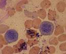 |
 | Polychromatic normoblast (also polychromatophilic) |  |
 | Orthochromatic normoblast (also orthochromatophilic) |  |
Pathosis
A megaloblast is an unusually large erythroblast that can be associated with vitamin B12 deficiency (caused by pernicious anemia or dietary insufficiency), folic acid deficiency, or both (such anemias are collectively called megaloblastic anemias). This kind of anemia leads to macrocytes (abnormally large red cells) and the condition called macrocytosis. The cause of this cellular gigantism is an impairment in DNA replication that delays nuclear maturation and cell division. Because RNA and cytoplasmic elements are synthesized at a constant rate despite the cells' impaired DNA synthesis, the cells show nuclear-cytoplasmic asynchrony.
Additional images
-

Blood cell lineage
-
Hematopoiesis
See also
References
- ↑ Hartenstein, V (2006). "Blood cells and blood cell development in the animal kingdom." (PDF). Annu Rev Cell Dev Biol. 22: 677–712. doi:10.1146/annurev.cellbio.22.010605.093317. Retrieved October 28, 2016.
- ↑ Osborn, Liz. "Number of Species Identified on Earth". www.currentresults.com. Current Results Publishing Ltd. Retrieved October 28, 2016.
- ↑ Schwarz, SO; Stansbury, F (1954). "Significance of nucleated red blood cells in peripheral blood: Analysis of 1,496 cases". JAMA. 154 (16): 1339–1340. doi:10.1001/jama.1954.02940500019007.
- ↑ Blood Smear: Details on RBCs, WBCs
- ↑ "Erythroblast" at Dorland's Medical Dictionary
- ↑ "Normoblast" at Dorland's Medical Dictionary
- ↑ "Megaloblast" at Dorland's Medical Dictionary
- ↑ Normoblasts at the US National Library of Medicine Medical Subject Headings (MeSH)
External links
- Pronormoblasts Presented by the University of Virginia
- Basophilic Normoblasts Presented by the University of Virginia
- Polychromatophilic Normoblasts Presented by the University of Virginia
- Orthochromatic Normoblasts Presented by the University of Virginia
- Histology image: 01804loa – Histology Learning System at Boston University - "Bone Marrow and Hemopoiesis bone marrow smear, erythroblast series with proerythroblast "
- Histology at uiowa.edu