Femoral artery
| Femoral artery | |
|---|---|
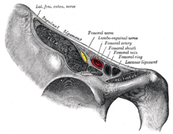 Structures passing behind the inguinal ligament. (Femoral artery labeled at upper right.) | |
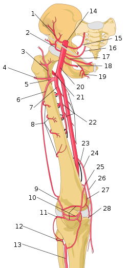 Schema of femoral artery (labeled as #20) and its major branches - right thigh, anterior view. | |
| Details | |
| Source | External iliac artery |
| Branches | Superficial epigastric artery, superficial iliac circumflex, superficial external pudendal, deep external pudendal, deep femoral artery, continues as popliteal artery |
| Vein | Femoral vein |
| Supplies | Anterior compartment of thigh |
| Identifiers | |
| Latin | Arteria femoralis |
| MeSH | A07.231.114.351 |
| TA | A12.2.16.010 |
| FMA | 70248 |
The femoral artery (Latin: arteria femoralis) is a large artery in the thigh and the main arterial supply to the lower limb. It enters the thigh from behind the inguinal ligament as the common femoral artery, a continuation of the external iliac artery. Here, it lies midway between the anterior superior iliac spine and the symphysis pubis. The common femoral artery gives off the profunda femoris artery and becomes the superficial femoral artery to descend along the anteromedial part of the thigh in the femoral triangle. It enters and passes through the adductor (subsartorial) canal, and becomes the popliteal artery as it passes through an opening in adductor magnus near the junction of the middle and distal thirds of the thigh.[1]
Structure
Its first three or four centimetres are enclosed, with the femoral vein, in the femoral sheath. The relations of the femoral artery are as follows:
- Anteriorly: In the upper part of its course, it is superficial and is covered by skin and fascia. In the lower part of its course, it passes behind the sartorius muscle.
- Posteriorly: The artery lies on the psoas, which separates it from the hip joint, the pectineus, and the adductor longus. The femoral vein intervenes between the artery and the adductor longus.
- Medially: It is related to the femoral vein in the upper part of its course.
- Laterally: The femoral nerve and its branches.
The femoral artery gives off several branches in the thigh which include;
- The superficial circumflex iliac artery is a small branch that runs up to the region of the anterior superior iliac spine.
- The superficial epigastric artery is a small branch that crosses the inguinal ligament and runs to the region of the umbilicus.
- The superficial external pudendal artery is a small branch that runs medially to supply the skin of the scrotum (or labium majus).
- The deep external pudendal artery runs medially and supplies the skin of the scrotum (or labium majus).
- The profunda femoris artery is a large and important branch that arises from the lateral side of the femoral artery about 1.5 in. (4 cm) below the inguinal ligament. It passes medially behind the femoral vessels and enters the medial fascial compartment of the thigh. It ends by becoming the fourth perforating artery. At its origin, it gives off the medial and lateral femoral circumflex arteries, and during its course it gives off three perforating arteries.
- The descending genicular artery is a small branch that arises from the femoral artery near its termination within the adductor canal. It assists in supplying the knee joint.
In clinical parlance, the part of the femoral artery proximal to the origin of profunda femoris is often termed the common femoral artery, while that distal to the origin of the profunda is termed the superficial femoral artery.[2]
Clinical significance
Pulse
As the femoral artery can often be palpated through the skin, it is often used as a catheter access artery. From it, wires and catheters can be directed anywhere in the arterial system for intervention or diagnostics, including the heart, brain, kidneys, arms and legs. The direction of the needle in the femoral artery can be against blood flow (retro-grade), for intervention and diagnostic towards the heart and opposite leg, or with the flow (ante-grade or ipsi-lateral) for diagnostics and intervention on the same leg. Access in either the left or right femoral artery is possible and depends on the type of intervention or diagnostic.
The site for optimally palpating the femoral pulse is in the inner thigh, at the mid-inguinal point, halfway between the pubic symphysis and anterior superior iliac spine. Presence of a femoral pulse has been estimated to indicate a systolic blood pressure of more than 50 mmHg, as given by the 50% percentile.[3]
The femoral artery can be used to draw arterial blood when the blood pressure is so low that the radial or brachial arteries cannot be located.
Peripheral arterial disease
The femoral artery is susceptible to peripheral arterial disease.[4] When it is blocked through atherosclerosis, percutaneous intervention with access from the opposite femoral may be needed. Endarterectomy, a surgical cut down and removal of the plaque of the femoral artery is also common. If the femoral artery has to be ligated surgically to treat a popliteal aneurysm, blood can still reach the popliteal artery distal to the ligation via the genicular anastomosis. However, if flow in the femoral artery of a normal leg is suddenly disrupted, blood flow distally is rarely sufficient. The reason for this is the fact that the genicular anastomosis is only present in a minority of individuals and is always undeveloped when disease in the femoral artery is absent.[5]
History
Textbook illustrations of the genicular anastomosis, such as that shown in the sidebox, all appear to have been derived from the idealized image first produced by Gray's Anatomy in 1910. Neither the 1910 illustration, nor any subsequent version, was made of an anatomical dissection but rather from the writings of John Hunter (surgeon) and Astley Cooper which described the genicular anastomosis many years after ligation of the femoral artery for popliteal aneurysm.[5] The genicular anastomosis has not been demonstrated even with modern imaging techniques such as X-ray computed tomography or angiography.[5]
See also
Brachial artery an arm based artery with a similar function
References
- ↑ Schulte, Erik; Schumacher, Udo (2006). "Arterial Supply to the Thigh". In Ross, Lawrence M.; Lamperti, Edward D. Thieme Atlas of Anatomy: General Anatomy and Musculoskeletal System. Thieme. p. 490. ISBN 978-3-13-142081-7.
- ↑ S. Snell, Richard (2008). Clinical Anatomy By Regions (8 ed.). Baltimore: Lippincott Williams & Wilkins. pp. 581–582.
- ↑ Deakin CD, Low JL (September 2000). "Accuracy of the advanced trauma life support guidelines for predicting systolic blood pressure using carotid, femoral, and radial pulses: observational study". BMJ. 321 (7262): 673–4. doi:10.1136/bmj.321.7262.673. PMC 27481
 . PMID 10987771.
 - ↑ Macpherson DS, Evans DH, Bell PR (January 1984). "Common femoral artery Doppler wave-forms: a comparison of three methods of objective analysis with direct pressure measurements". The British Journal of Surgery. 71 (1): 46–9. doi:10.1002/bjs.1800710114. PMID 6689970.
- 1 2 3 Sabalbal M, Johnson M, McAlister V (September 2013). "Absence of the genicular arterial anastomosis as generally depicted in textbooks". Annals of the Royal College of Surgeons of England. 95 (6): 405–9. doi:10.1308/003588413X13629960046831. PMID 24025288.
Additional images
 Structures surrounding right hip-joint.
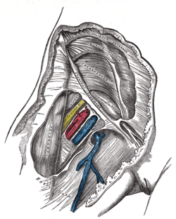 Femoral sheath laid open to show its three compartments.
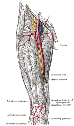 The femoral artery.
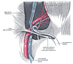 The spermatic cord in the inguinal canal.
 Front of right thigh, showing surface markings for bones, femoral artery and femoral nerve.
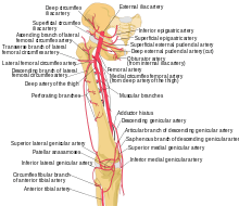 Schema of arteries of the thigh.
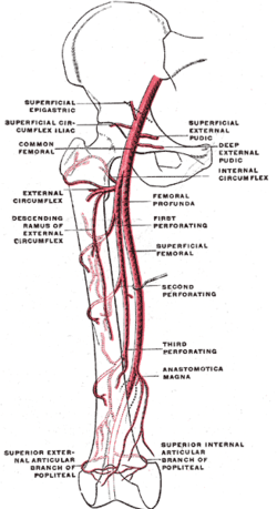 Femoral artery and its major branches - right thigh, anterior view.
 Illustration depicting main leg arteries (anterior view).
 Femoral artery - deep dissection.
 Femoral artery - deep dissection.
External links
| Wikimedia Commons has media related to Femoral artery. |
- Anatomy photo:12:05-0101 at the SUNY Downstate Medical Center
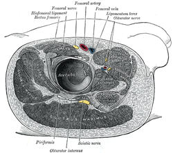
- Cross section image: pelvis/pelvis-e12-15 - Plastination Laboratory at the Medical University of Vienna
- Image at umich.edu - pulse
- Diagram at MSU