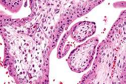
Hofbauer cell
Hofbauer cells are oval eosinophilic histiocytes[1] with granules and vacuoles found in the placenta, which are of mesenchymal origin, in mesoderm of the chorionic villus, particularly numerous in early pregnancy.
Etymology
They are named after J. Isfred Isidore Hofbauer, an American gynecologist. (1878-1961)[1]
Function
They are believed to be a type of macrophage[2][3] and are most likely involved in preventing the transmission of pathogens from the mother to the fetus (so-called vertical transmission). Although there are many studies concerning placental vasculogenesis and angiogenesis, there has been a lack of evidence on the possible roles of Hofbauer cells in these processes.[4]
Histology

Under histology sections, Hofbauer cells have appeared with discernible amount of cytoplasm.
See also
References
- 1 2 Venes, Donald (2006). Taber's cyclopedic medical dictionary (Ed. 20, illustrated in full color. ed.). Philadelphia [Pa.]: Davis Co. ISBN 0-8036-1208-7.
- ↑ Wood, GW. "Mononuclear phagocytes in the human placenta.". Placenta. 1 (2): 113–23. doi:10.1016/s0143-4004(80)80019-1. PMID 7003580.
- ↑ Zaccheo, D.; Pistoia, V.; Castellucci, M.; Martinoli, C. (1989). "Isolation and characterization of Hofbauer cells from human placental villi.". Arch Gynecol Obstet. 246 (4): 189–200. doi:10.1007/bf00934518. PMID 2482706.
- ↑ Seval, Y.; Korgun, ET.; Demir, R. "Hofbauer cells in early human placenta: possible implications in vasculogenesis and angiogenesis.". Placenta. 28 (8-9): 841–5. doi:10.1016/j.placenta.2007.01.010. PMID 17350092.