KaiC
kaiC is a gene belonging to the kaiABC gene cluster. The kaiABC gene cluster is a group of genes, consisting of kaiA, kaiB, and kaiC, globally regulating bacterial circadian rhythms, specifically in cyanobacteria. Regulation of kaiABC expression is essential for circadian rhythmicity, and is particularly important for regulating cyanobacteria processes such as nitrogen fixation, photosynthesis, and cell division.[1] Using bacterial luciferase as a reporter for gene expression, studies have shown that, similar to Drosophila, mouse, and Neurospora clock models, the cyanobacteria circadian clock, controlled by kaiABC regulation, is also based on a transcription translation feedback loop(TTFL).[2] Within its gene cluster, kaiC codes for the regulator, KaiC protein, which not only suppresses kaiBC when overexpressed, but also suppresses circadian expression of all genes in the cyanobacterial genome.[3]
| kaiC | |
|---|---|
| Identifiers | |
| Organism | |
| Symbol | kaiC |
| Entrez | 3773504 |
| RefSeq (Prot) | YP_400233.1 |
History
Though the kaiABC gene cluster has been found to exist only in cyanobacteria, evolutionarily kaiC contains
homologs that occur in Archaea and Proteobacteria. It is the oldest circadian gene that has been discovered in prokaryotes. kaiC has a double-domain structure and sequence that classifies it as part of the RecA gene family of ATP-dependent recombinases.[1] Based on a number of single-domain homologous genes in other species, kaiC is hypothesized to have horizontally transferred from Bacteria to Archaea, eventually forming the double-domain kaiC through duplication and fusion. kaiC's key role in circadian control and homology to RecA suggest its individual evolution before its presence in the kaiABC gene cluster.[4]
Discovery
Pioneers Takao Kondo, Susan S. Golden, and Carl H. Johnson discovered the gene cluster in 1998 and named the gene cluster kaiABC, as "kai" means “cycle” in Japanese. They generated 19 different clock mutants that were mapped to kaiA, kaiB, and kaiC genes, and successfully cloned the gene cluster in the cyanobacteria Synechococcus elongatus. Using a bacterial luciferase reporter to monitor the expression of clock-controlled gene psbAI in Synechococcus, they investigated and reported on the rescue to normal rhythmicity of long-period clock mutant C44a (with a period of 44 hours) by kaiABC. They inserted wild-type DNA through a pNIBB7942 plasmid vector into the C44a mutant, and generated clones that restored normal period (a period of 25 hours). They were eventually able to localize the gene region causing this rescue, and observed circadian rhythmicity in upstream promotor activity of kaiA and kaiB, as well as in the expression of kaiA and kaiBC messenger RNA. They determined abolishing any of the three kai genes would cause arrhythmicity in the circadian clock and reduce kaiBC promoter activity.[1]
Genetics and protein structure
On Synechococcus elongatus' singular circular chromosome, the protein-coding gene kaiC is located at position 380696-382255 (its locus tag is syc0334_d). The gene kaiC has paralogs kaiB (located 380338..380646) and kaiA (located 379394..380248). kaiC encodes the protein KaiC (519 amino acids). KaiC acts as a non-specific transcription regulator that represses transcription of the kaiBC promoter. Its crystal structure has been solved at 2.8 Å resolution; it is a homohexameric complex (approximately 360 kDa) with a double-doughnut structure and a central pore which is open at the N-terminal ends and partially sealed at the C-terminal ends due to the presence of six arginine residues.[5] The hexamer has twelve ATP molecules between the N- (CI) and C-terminal (CII) domains, which demonstrate ATPase activity.[6] Interfaces on KaiC's CII domain are sites for both auto-kinase and auto-phosphatase activity, both in vitro and in vivo.[7][8] KaiC has two P loops or Walker’s motif As (ATP-/GTP-binding motifs) in the CI and CII domains; the CI domain also contains two DXXG (X represents any amino acid) motifs that are highly conserved among the GTPase super-family.[9]
Function
Cyanobacteria are the simplest organisms for which a mechanism is known for the generation of circadian rhythms.[10] KaiC is temperature compensated from 25 to 50 degrees Celsius [11] and has a Q10 of about 1.1. Because the period of KaiC phosphorylation is temperature compensated and agrees with in vivo circadian rhythms, KaiC is thought to be the mechanism for basic circadian timing in Synechococcus.[12] ∆kaiABC individuals, one of the more common mutants, grow just as well as wild type individuals but lack rhythmicity. This is evidence that the kaiABC gene cluster is not necessary for growth.[5]
Regulation of KaiC
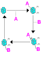
Kai proteins regulate genome-wide gene expression.[13] Protein KaiA enhances the phosphorylation of protein KaiC through control of the autophosphatase activity, while protein KaiB attenuates the activity of KaiA.[8] Disruption of KaiC’s CI domain results both in arrhythmia of kaiBC expression and a reduction of ATP-binding activity; this, along with in vitro autophosphorylation of KaiC indicate that ATP binding to KaiC is crucial for Synechococcus circadian oscillation.[9] The phosphorylation status of KaiC has been correlated with Synechococcus clock speed in vivo.[14] Additionally, overexpression of KaiC has been shown to strongly repress the kaiBC promoter, while kaiA overexpression has experimentally enhanced the kaiBC promoter.[5] These positive and negative binding elements mirror a feedback mechanism of rhythm generation conserved across many different species.[15] Phosphorylation of KaiC at subunits occurs in an ordered manner, beginning with phosphorylation of Threonine 432 (T432) followed by Serine 431 (S431), followed up by dephosphorylation in the same order.[16]
KaiC phosphorylation oscillates with a period of approximately 24 hours when placed in vitro with the three recombinant Kai proteins, incubated with ATP. The circadian rhythm of KaiC phosphorylation persists in constant darkness, regardless of Synechococcus transcription rates. This oscillation rate is thought to be controlled by the ratio of phosphorylated to unphosphorylated KaiC protein. KaiC phosphorylation ratio is a main factor in the activation of kaiBC promoter as well. The kaiBC operon is transcribed in a circadian fashion and precedes KaiC build up by about 6 hours,[10] a lag thought to play a role in feedback loops.
Interdependence of Kai A, B, and C
kaiA, kaiB, and kaiC have been shown to be essential genetic components in Synechococcus elongatus for circadian rhythms.[10] Experiments have also shown that KaiC enhances the KaiA-KaiB interaction in yeast cells and in vitro. This implies that there may be the formation of a heteromultimeric complex composed of the three Kai proteins with KaiC serving as a bridge between KaiA and KaiB. Alternatively, KaiC may form a heterodimer with KaiA or KaiB to induce a conformational change.[17] Variations in the C-terminal region of each of their proteins suggest functional divergence between the Kai clock proteins,[13] however there are critical interdependencies between the three paralogs.
Circadian Regulation of Cell Division
Recent experiments have found that the oscillations in the cell cycle and circadian rhythms of Synechococcus are linked together through a one way mechanism. The circadian clock gates cells division, only allowing it to proceed at certain phases. The cell cycle does not appear to have any effect on the circadian clock though. When binary fission occurs, the daughter cells inherit the mother cell's circadian clock and are in phase with the mother cell. The circadian gating of cell division may be a protective feature to prevent division at a vulnerable phase. Phases in which KaiC has high ATPase activity do not allow for cell division to take place. In mutants with constantly elevated KaiC ATPase activity, the protein CikA is absent. CikA is a major factor in the input pathway and causes KaiC dependent cell elongation.[18]
Notable research
The recreation of a circadian oscillator in vitro in the presence of only KaiA, KaiB, KaiC, and ATP has sparked interest in the relationship between cellular biochemical oscillators and their associated transcription-translation feedback loops (TTFLs). TTFLs have long been assumed to be the core of circadian rhythmicity, but that claim is now being tested again due to the possibility that the biochemical oscillators could constitute the central mechanism of the clock system, regulating and operating within TTFLs that control output and restore proteins essential to the oscillators in organisms, such as the KaiABC system in Synechococcus.[2] Two models have been proposed to describe the relationship between the biochemical and TTFL regulation of circadian rhythms: a master/slave oscillator system with the TTFL oscillator synchronizing to the biochemical oscillator and an equally weighted coupled oscillator system in which both oscillators synchronize and influence the other oscillator. Both are coupled oscillator models that account for the high stability of the timing mechanism within Synechococcus. The biochemical oscillator relies on redundant molecular interactions based on the law of mass action, whereas the TTFL relies on cellular machinery that mediates translation, transcription, and degradation of mRNA and proteins. The different types of interactions driving the two oscillators allows the circadian clock to be resilient to changes within the cell, such as metabolic fluctuation, temperature changes, and cell division.[19]
Though the period of the circadian clock is temperature compensated, the phosphorylation of KaiC can be stably entrained to a temperature cycle. The phosphorylation of KaiC was successfully entrained in vitro to temperature cycles with periods between 20 and 28 hours using temperature steps from 30 °C to 45 °C and vice versa. The results reflect a phase-dependent shift in the phase of the KaiC phosphorylation rhythms. The period of the circadian clock was not changed, reinforcing the temperature compensation of the clock mechanism.[20]
A 2012 study out of Vanderbilt University shows evidence that KaiC acts as a phospho-transferase that hands back phosphates to ADP on the T432 (threonine residue at position 432) and S431 (serine residue 431) indicating that KaiC effectively serves as an ATP synthase.[6]
Various KaiC mutants have been identified and their phenotypes studied. Many mutants show a change in the period of their circadian rhythms.
| Mutation | Period |
|---|---|
| Wild | 24.8 Hours |
| E318A | Arrhythmic |
| E318D | Arrhythmic |
| R385A | 36-48 Hours |
| D417A | 25.6 Hours |
| H429A | 28.0 Hours |
| I430A | Arrhythmic |
| F470Y | 17 Hours |
| S157P | 21 Hours |
| T42S | 28 Hours |
See also
References
- 1 2 3 Ishiura, M; Kutsuna, S; Aoki, S; Iwasaki, H; Andersson, C. "R, Tanabe A, Golden S S, Johnson C H, Kondo T. (1998)". Science. 281: 1519–1523.
- 1 2 Egli, M; Johnson, CH (2013). "A circadian clock nanomachine that runs without transcription or translation". Curr. Opin. Neurobiol. 23: 732–740. doi:10.1016/j.conb.2013.02.012.
- ↑ Nakahira, Y.; Katayama, M.; Miyashita, H.; Kutsuna, S.; Iwasaki, H.; Oyama, T.; Kondo, T. (2004). "Global gene repression by KaiC as a master process of prokaryotic circadian system". PNAS. 101 (3): 881–885. doi:10.1073/pnas.0307411100.
- ↑ Dvornyk, V (2003). "Origin and evolution of circadian clock genes in prokaryotes". PNAS. 100: 2495–2500. doi:10.1073/pnas.0130099100.
- 1 2 3 Ishiura, M. 1998. Expression of a gene cluster kaiABC as a circadian feedback process in cyanobacteria. Science.
- 1 2 Egli, M.; Mori, T.; Pattanayek, R.; Xu, Y.; Qin, X.; Johnson, C. H. (2012). "Dephosphorylation of the core clock protein KaiC in the cyanobacterial KaiABC circadian oscillator proceeds via an ATP synthase mechanism". Biochemistry. 51 (8): 1547–1558. doi:10.1021/bi201525n.
- ↑ Iwasaki, Hideo et al. KaiA-Stimulated KaiC Phosphorylation in Circadian Timing Loops in Cyanobateria. Proceedings of the National Academy of Sciences of the United States of America 99.24 (2002): 15788-15793. PMC. Web. 9 Apr. 2015.
- 1 2 Xu, Y.; Mori, T.; Johnson, C. H. (2003). "Cyanobacterial circadian clockwork: roles of KaiA, KaiB and the kaiBC promoter in regulating KaiC". EMBO J. 22: 2117–2126. doi:10.1093/emboj/cdg168.
- 1 2 Nishiwaki, T; Iwasaki, H; Ishiura, M; Kondo (2000). "Nucleotide binding and autophosphorylation of the clock protein KaiC as a circadian timing process of cyanobacteria". Proc Natl Acad Sci U S A. 97: 495–499. doi:10.1073/pnas.97.1.495.
- 1 2 3 Murayama, Y.; Oyama, T.; Kondo, T. (2008). "Regulation of circadian clock gene expression by phosphorylation states of KaiC in cyanobacteria". Journal of Bacteriology. 190 (5): 1691–1698. doi:10.1128/jb.01693-07.
- ↑ Murakami, R.; Miyake, A.; Iwase, R.; Hayashi, F.; Uzumaki, T.; Ishiura, M. (2008). "ATPase activity and its temperature compensation of the cyanobacterial clock protein KaiC". Genes to Cells. 13 (4): 387–395. doi:10.1111/j.1365-2443.2008.01174.x.
- ↑ Terauchi, K.; Kitayama, Y.; Nishiwaki, T.; Miwa, K.; Murayama, Y.; Oyama, T.; Kondo, T. (2007). "ATPase activity of KaiC determines the basic timing for circadian clock of cyanobacteria". Proc. Natl. Acad. Sci. USA. 104: 16377–16381. doi:10.1073/pnas.0706292104.
- 1 2 Tomita, J.; Nakajima, M.; Kondo, T.; Iwasaki, H. (2005). "No transcription-translation feedback in circadian rhythm of KaiC phosphorylation". Science. 307: 251–254. doi:10.1126/science.1102540.
- ↑ Xu, Y.; Mori, T.; Johnson, C. (2003). "Cyanobacterial circadian clockwork: roles of KaiA, KaiB, and the "kaiBC" promoter in regulating KaiC". The EMBO Journal. 22: 2117–2126. doi:10.1093/emboj/cdg168.
- ↑ Dunlap, Jay C (1999). "Molecular Bases for Circadian Clocks". Cell. 96 (2): 271–90. doi:10.1016/s0092-8674(00)80566-8.
- ↑ Phong, Connie; et al. (2013). "Robust and Tunable Circadian Rhythms from Differentially Sensitive Catalytic Domains". Proceedings of the National Academy of Sciences of the United States of America. 110 (3): 1124–1129. doi:10.1073/pnas.1212113110.
- ↑ Iwasaki, H.; Taniguchi, Y.; Ishiura, M.; Kondo, T. (1999). "Physical interactions among circadian clock proteins KaiA, KaiB and KaiC in cyanobacteria". The EMBO Journal. 18 (5): 1137–1145. doi:10.1093/emboj/18.5.1137.
- ↑ Dong, Guogang; Kim, Yong-Ick; Golden, Susan S. "Simplicity and complexity in the cyanobacterial circadian clock mechanism". Current Opinion in Genetics & Development. 20 (6): 619–625. doi:10.1016/j.gde.2010.09.002.
- ↑ Johnson, Carl Hirschie; Egli, Martin (2014). "Metabolic Compensation and Circadian Resilience in Prokaryotic Cyanobacteria". Annual Review of Biochemistry. 83: 221–247. doi:10.1146/annurev-biochem-060713-035632.
- ↑ Yoshida, Takuya; et al. (2009). "Nonparametric Entrainment of the in Vitro Circadian Phosphorylation Rhythm of Cyanobacterial KaiC by Temperature Cycle". Proceedings of the National Academy of Sciences of the United States of America. 106 (5): 1648–1653. doi:10.1073/pnas.0806741106.
- ↑ Nakajima, Masato; et al. (2005). "Reconstitution of Circadian Oscillation of Cyanobacterial KaiC Phosphorylation in Vitro". Science. 308 (5720): 414–415. doi:10.1126/science.1108451.
- ↑ Pattanayek, Rekha; et al. (2009). "Structures of KaiC Circadian Clock Mutant Proteins: A New Phosphorylation Site at T426 and Mechanisms of Kinase, ATPase and Phosphatase". PLoS ONE. 4 (11): e7529. doi:10.1371/journal.pone.0007529.