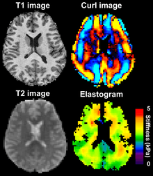
Magnetic resonance elastography

Magnetic resonance elastography (MRE) is a non-invasive medical imaging technique that measures the mechanical properties (stiffness) of soft tissues by introducing shear waves and imaging their propagation using MRI.[1] Pathological tissues are often stiffer than the surrounding normal tissue. For instance, malignant breast tumors are much harder than healthy fibro-glandular tissue. This characteristic has been used by physicians for screening and diagnosis of many diseases, through palpation. MRE calculates the mechanical parameter as elicited by palpation, in a non-invasive and objective way.
Magnetic resonance elastography works by using an additional gradient waveform in the pulse sequence to sensitize the MRI scan to shear waves in the tissue. The shear waves are generated by an electro-mechanical transducer on the surface of the skin. Both the mechanical excitation and the motion sensitizing gradient are at the same frequency. This encodes the amplitude of the shear wave in the tissue in the phase of the MRI image. An algorithm can be used to extract a quantitative measure of tissue stiffness from the MRI in an elastogram.
Magnetic resonance elastography (MRE) was pioneered by researchers at the Mayo Clinic, the University of Michigan, and the University of Toronto, starting from the mid 1990s.[2][3][4][5] MRE is being investigated to be used for a multitude of diseases that affect the tissue stiffness.[6] It is currently being clinically used for the assessment of hepatic fibrosis,[7][8][9] since it is well known that the liver stiffness increases with the progression of this disease. It is being investigated for the diagnosis of diseases and also for studying the treatment efficacy. For instance, this has been utilized in ablative treatments using high-intensity focused ultrasound where the treated necrosed tissue can be distinguished with MRE even in real time.
References
- ↑ Muthupillai R; Lomas, DJ; Rossman, PJ; Greenleaf, JF; Manduca, A; Ehman, RL (1995). "Magnetic resonance elastography by direct visualization of propagating acoustic strain waves". Science. 269 (5232): 1854–1857. doi:10.1126/science.7569924. PMID 7569924.
- ↑ Sarvazyan A, Hall TJ, Urban MW, Fatemi M, Aglyamov SR, Garra BS. An overview of elastography - an emerging branch of medical imaging. Current medical imaging reviews 2011; 7 (4): 255–282.
- ↑ Sarvazyan AP, Skovoroda AR, Emelianov SY, Fowlkes JB, Pipe JG, Adler RS, Buxton RB, Carson PL. Biophysical bases of elasticity imaging. In: Acoustical Imaging. Ed. Jones JP, Plenum Press, New York and London, 1995; 21: 223-240.
- ↑ Plewes DB, Betty I, Urchuk SN, and Soutar I, Visualizing tissue compliance with MR imaging. J. Magn. Res. Imaging 1995; 5: 733-738.
- ↑ Fowlkes JB, Emelianov SY, Pipe JG, Skovoroda AR, Carson PL, Adler RS, and Sarvazyan AP, Magnetic-resonance imaging techniques for detection of elasticity variation. Med. Phys. 1995; 22: 1771-8.
- ↑ Mariappan YK; Glaser, KJ; Ehman, RL (2010). "Magnetic resonance elastography: A review". Clinical Anatomy. 23 (5): 497–511. doi:10.1002/ca.21006.
- ↑ Meng Yin; Talwalker, JA; Glaser, KJ; Manduca, A; Grimm, R; Rossman, P; Fidler, JL; Ehman, RL (2007). "Assessment of Hepatic Fibrosis with Magnetic resonance elastography". Clinical Gastroenterology and Hepatology. 5 (10): 1207–1213. doi:10.1016/j.cgh.2007.06.012.
- ↑ Huwart L; Sempoux, C; Vicaut, E; Salameh, N; Laurence, A; Danse, E; Peeters, F; Beek, L; Rahier, J; Sinkus, R; Horsmans, Yves; Beers, BE (2008). "Magnetic Resonance Elastography for the noninvasive staging of liver fibrosis". Gastroenterology. 135 (1): 32–40. doi:10.1053/j.gastro.2008.03.076.
- ↑ Asbach P; Klatt, D; Schlosser, B; Biermer, M; Muche, M; Rieger, A; Loddenkemper, C; Somasundaram, R; Berg, T; Hamm, B; Braun, J; Sack, I (2010). "Viscoelasticity-based Staging of Hepatic Fibrosis with Multifrequency MR Elastography". Radiology. 257 (1): 80–86. doi:10.1148/radiol.10092489.
See also
| Wikimedia Commons has media related to Magnetic resonance elastography. |