Orbital process of the zygomatic bone
| Orbital process of the zygomatic bone | |
|---|---|
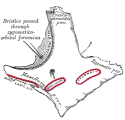
 Left zygomatic bone. Malar surface. (Orbital proc. visible at upper left.) | |
| Details | |
| Identifiers | |
| Latin | facies orbitalis ossis zygomatici |
| TA | A02.1.14.004 |
| FMA | 59226 |
The orbital process of the zygomatic bone is a thick, strong plate, projecting backward and medialward from the orbital margin.
Its antero-medial surface forms, by its junction with the orbital surface of the maxilla and with the great wing of the sphenoid, part of the floor and lateral wall of the orbit.
On it are seen the orifices of two canals, the zygomatico-orbital foramina; one of these canals opens into the temporal fossa, the other on the malar surface of the bone; the former transmits the zygomaticotemporal, the latter the zygomaticofacial nerve.
- Its postero-lateral surface, smooth and convex, forms parts of the temporal and infratemporal fossae.
- Its anterior margin, smooth and rounded, is part of the circumference of the orbit.
- Its superior margin, rough, and directed horizontally, articulates with the frontal bone behind the zygomatic process.
- Its posterior margin is serrated for articulation, with the great wing of the sphenoid and the orbital surface of the maxilla.
At the angle of junction of the sphenoidal and maxillary portions, a short, concave, non-articular part is generally seen; this forms the anterior boundary of the inferior orbital fissure: occasionally, this non-articular part is absent, the fissure then being completed by the junction of the maxilla and sphenoid, or by the interposition of a small sutural bone in the angular interval between them.
See also
Additional images
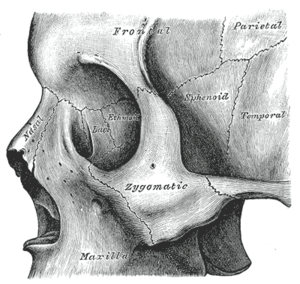
 Left zygomatic bone in situ.
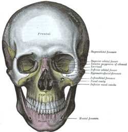
 The skull from the front.
 The seven bones which articulate to form the orbit.
References
This article incorporates text in the public domain from the 20th edition of Gray's Anatomy (1918)