Phototaxis
| Wikimedia Commons has media related to Phototaxis. |

Phototaxis is a kind of taxis, or locomotory movement, that occurs when a whole organism moves towards or away from stimulus of light.[1] This is advantageous for phototrophic organisms as they can orient themselves most efficiently to receive light for photosynthesis. Phototaxis is called positive if the movement is in the direction of increasing light intensity and negative if the direction is opposite.[2]
Two types of positive phototaxis are observed in prokaryotes. The first is called scotophobotaxis (from the word "scotophobia"), which is observed only under a microscope. This occurs when a bacterium swims by chance out of the area illuminated by the microscope. Entering darkness signals the cell to reverse flagella rotation direction and reenter the light. The second type of phototaxis is true phototaxis, which is a directed movement up a gradient to an increasing amount of light. This is analogous to positive chemotaxis except that the attractant is light rather than a chemical.
Phototactic responses are observed in many organisms such as Serratia marcescens, Tetrahymena, and Euglena. Each organism has its own specific biological cause for a phototactic response, many of which are incidental and serve no end purpose.
Phototaxis in zooplankton
Phototaxis in zooplankton is well studied in the marine annelid Platynereis dumerilii:
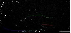
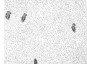
Platynereis dumerilii trochophore and metatrochophore larvae are positively phototactic. Phototaxis there is mediated by simple eyespots that consists of a pigment cell and a photoreceptor cell. The photoreceptor cell synapses directly onto ciliated cells, which are used for swimming. The eyespots do not give spatial resolution, therefore the larvae are rotating to scan their environment for the direction where the light is coming from.[4]
Platynereis dumerilii nectochate larvae can switch between positive and negative phototaxis. Phototaxis there is mediated by two pairs of more complex pigment cup eyes. These eyes contain more photoreceptor cells that are shaded by pigment cells forming a cup. The photoreceptor cells do not synapse directly onto ciliated cells or muscle cells but onto inter-neurons of a processing center. This way the information of all four eye cups can be compared and a low resolution image of four pixels can be created telling the larvae where the light is coming from. This way the larva does not need to scan its environment by rotating.[3] This is an adaption for living on the bottom of the sea the life style of the nectochaete larva while scanning rotation is more suited for living in the open water column, the life style of the trochophore larva. Phototaxis in the Platynereis dumerilii nectochate larva has a broad spectral range which is at least covered by three opsins that are expressed by the cup eyes:[5] Two rhabdomeric opsins[6] and a Go-opsin.[5]
See also
Further reading
- Madigan, Michael T.; Martinko, John M. (2006). Brock Biology of Microorganisms (11th ed.). Upper Saddle River, N.J.: Pearson/Prentice Hall. ISBN 0131443291.
- Jékely, G. (31 August 2009). "Evolution of phototaxis". Philosophical Transactions of the Royal Society B: Biological Sciences. 364 (1531): 2795–2808. doi:10.1098/rstb.2009.0072. PMC 2781859
 . PMID 19720645.
 - Randel, Nadine; Jékely, Gáspár (23 November 2015). "Phototaxis and the origin of visual eyes". Philosophical Transactions of the Royal Society B: Biological Sciences. 371 (1685): 20150042. doi:10.1098/rstb.2015.0042. PMID 26598725.
References
- ↑ Martin, E.A., ed. (1983), Macmillan Dictionary of Life Sciences (2nd ed.), London: Macmillan Press, p. 362, ISBN 0-333-34867-2
- ↑ Menzel, Randolf (1979), "Spectral Sensitivity and Color Vision in Invertebrates", in H. Autrum (editor), Comparative Physiology and Evolution of Vision in Invertebrates- A: Invertebrate Photoreceptors, Handbook of Sensory Physiology, VII/6A, New York: Springer-Verlag, pp. 503–580. See section D: Wavelength–Specific Behavior and Color Vision, ISBN 3-540-08837-7
- 1 2 3 Randel, Nadine; Asadulina, Albina; Bezares-Calderón, Luis A; Verasztó, Csaba; Williams, Elizabeth A; Conzelmann, Markus; Shahidi, Réza; Jékely, Gáspár (27 May 2014). "Neuronal connectome of a sensory-motor circuit for visual navigation". eLife. 3. doi:10.7554/eLife.02730.
- ↑ Jékely, Gáspár; Colombelli, Julien; Hausen, Harald; Guy, Keren; Stelzer, Ernst; Nédélec, François; Arendt, Detlev (20 November 2008). "Mechanism of phototaxis in marine zooplankton". Nature. 456 (7220): 395–399. doi:10.1038/nature07590.
- 1 2 Gühmann, Martin; Jia, Huiyong; Randel, Nadine; Verasztó, Csaba; Bezares-Calderón, Luis A.; Michiels, Nico K.; Yokoyama, Shozo; Jékely, Gáspár (August 2015). "Spectral Tuning of Phototaxis by a Go-Opsin in the Rhabdomeric Eyes of Platynereis". Current Biology. 25 (17): 2265–2271. doi:10.1016/j.cub.2015.07.017.
- ↑ Randel, N.; Bezares-Calderon, L. A.; Gühmann, M.; Shahidi, R.; Jekely, G. (10 May 2013). "Expression Dynamics and Protein Localization of Rhabdomeric Opsins in Platynereis Larvae". Integrative and Comparative Biology. 53 (1): 7–16. doi:10.1093/icb/ict046.