Placentation
| Placentation | |
|---|---|
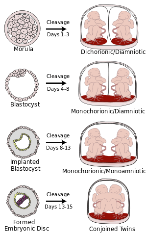 Placentation resulting from cleavage at various gestational ages | |
| Details | |
| Latin | placentatio |
In biology, placentation refers to the formation, type and structure, or arrangement of placentas. The function of placentation is to transfer nutrients from maternal tissue to a growing embryo. Placentation is best known in pregnant female mammals (eutheria), but also occurs in other animals, eggs (yolk sac placentation) and flowering plants.
Placentation in mammals
In placental mammals, the placenta forms after the embryo implants into the wall of the uterus. The developing fetus is connected to it via an umbilical cord. Animal placentas are classified based on the number of tissues separating the maternal from the fetal blood. The placentation types found in animals are:
- endotheliochorial placentation
In this type of placentation, the chorionic villi are in contact with the endothelium of maternal blood vessels. (e.g. in most carnivores like cats and dogs)
- epitheliochorial placentation
Chorionic villi, growing into the apertures of uterine glands ( epithelium). (e.g. in ruminants, horses, whales, lower primates)
- hemochorial placentation (e.g. in higher order primates, including humans, and also in rabbits, guinea pigs, mice, and rats)[1]
In hemochorial placentation maternal blood comes in direct contact with the fetal chorion, which it does not in the other two types.[2] It may avail for more efficient transfer of nutrients etc., but is also more challenging for the systems of gestational immune tolerance to avoid rejection of the fetus.[3]
During pregnancy, placentation is the formation and growth of the placenta inside the uterus. It occurs after the implantation of the embryo into the uterine wall and involves the remodeling of blood vessels in order to supply the needed amount of blood. In humans, placentation takes place 7–8 days after fertilization.
In humans, the placenta develops in the following manner. Chorionic villi (from the embryo) on the embryonic pole grow, forming chorion frondosum. Villi on the opposite side (abembryonic pole) degenerate and form the chorion laeve (or chorionic laevae), a smooth surface. The endometrium (from the mother) over the chorion frondosum (this part of the endometrium is called the decidua basalis) forms the decidual plate. The decidual plate is tightly attached to the chorion frondosum and goes on to form the actual placenta. Endometrium on the opposite side to the decidua basalis is the decidua parietalis. This fuses with the chorion laevae, thus filling up the uterine cavity.[4]
In the case of twins, dichorionic placentation refers to the presence of two placentas (in all dizygotic and some monozygotic twins). Monochorionic placentation occurs when monozygotic twins develop with only one placenta and bears a higher risk of complications during pregnancy. Abnormal placentation can lead to an early termination of pregnancy, for example in pre-eclampsia.
Placentation in lizards and snakes
As placentation is essential for live birth, the more than 100 origins of live birth in lizards and snakes (Squamata) have seen an equal number of independent origins of placentation. This means that the occurrence of placentation in squamata is more frequent than in all other vertebrates combined,[5] making them ideal for research on the evolution of placentation and viviparity itself. In most squamates two separate placentae form, utilising separate embryonic tissue (the chorioallantoic and yolk-sac placentae). In species with more complex placentation, we see regional specialisation for gas,[6] amino acid,[7] and lipid transport.[8] Placentae form following implantation into uterine tissue (as seen in mammals) and formation is likely facilitated by a plasma membrane transformation.[9]
Most reptiles exhibit strict epitheliochorial placentation (e.g. Pseudemoia entrecasteauxii) however at least two examples of endotheliochorial placentation have been identified (Mabuya sp. and Trachylepis ivensi).[10] However, unlike eutherian mammals, epitheliochorial placentation is not maintained by maternal tissue as embryos do not readily invade maternal tissue outside of the uterus.[11]
Placentation in plants
In flowering plants, placentation occurs where the ovules are attached inside the ovary.[12] The ovules inside a flower's ovary (which later become the seeds inside a fruit) are attached via funiculi, the plant part equivalent to an umbilical cord. The part of the ovary where the funiculus attaches is referred to as the placenta.
In botany, the term placentation most commonly refers to the arrangement of placentas inside a flower or fruit. Plant placentation types include:
- Basal placentation: The placenta is at the base (bottom) of the ovary. Simple or compound carpel.
- Apical placentation: The placenta is at the apex (top) of the ovary. Simple or compound carpel.
- Parietal placentation: The placentas are in the ovary wall within a non-sectioned ovary. Compound carpel.
- Axile placentation: The ovary is sectioned by radial spokes with placentas in separate locules. Compound carpel.
- Free or central placentation: The placentas are in a central column within a non-sectioned ovary. Compound carpel.
- Marginal placentation: There is only one elongated placenta on one side of the ovary, as ovules are attached at the fusion line of the carpel's margins . This is conspicuous in legumes. Simple carpel.
See also
References
- ↑ Claim for guinea pigs, rabbits, mice, and rats taken from: Thornburg KL, Faber JJ (October 1976). "The steady state concentration gradients of an electron-dense marker (ferritin in the three-layered hemochorial placenta of the rabbit". J. Clin. Invest. 58 (4): 912–25. doi:10.1172/JCI108544. PMC 333254
 . PMID 965495.
 - ↑ thefreedictionary.com > hemochorial placenta Citing: Dorland's Medical Dictionary for Health Consumers. Copyright 2007 by Saunders
- ↑ Elliot, M.; Crespi, B. (2006). "Placental invasiveness mediates the evolution of hybrid inviability in mammals". The American Naturalist. 168 (1): 114–120. doi:10.1086/505162. PMID 16874618.
- ↑ T.W. Sadler, Langman's Medical Embryology, 11th edition, Lippincott & Wilkins
- ↑ Blackburn, DG; Flemming, AF (2009). "Morphology, development, and evolution of fetal membranes and placentation in squamate reptiles". J. Exp. Zool. (Mol. Dev. Evol.). 312B: 579–589. doi:10.1002/jez.b.21234.
- ↑ Adams, S. M., Biazik, J. M., Thompson, M. B., & Murphy, C. R. (2005). Cyto‐epitheliochorial placenta of the viviparous lizard Pseudemoia entrecasteauxii: A new placental morphotype. Journal of morphology, 264(3), 264-276.Chicago
- ↑ Itonaga, K., Wapstra, E., & Jones, S. M. (2012). A novel pattern of placental leucine transfer during mid to late gestation in a highly placentotrophic viviparous lizard. Journal of Experimental Zoology Part B: Molecular and Developmental Evolution, 318(4), 308-315.
- ↑ Griffith, O. W., Ujvari, B., Belov, K., & Thompson, M. B. (2013). Placental lipoprotein lipase (LPL) gene expression in a placentotrophic lizard, Pseudemoia entrecasteauxii. Journal of Experimental Zoology Part B: Molecular and Developmental Evolution.
- ↑ Murphy, C. R.; Hosie, M. J.; Thompson, M. B. (2000). "The plasma membrane transformation facilitates pregnancy in both reptiles and mammals". Comparative Biochemistry and Physiology A. 127 (4): 433–439. doi:10.1016/s1095-6433(00)00274-9.
- ↑ Blackburn, D. G., & Flemming, A. F. (2010). Reproductive specializations in a viviparous African skink: Implications for evolution and biological conservation.
- ↑ Griffith, O. W.; Van Dyke, J. U.; Thompson, M. B. (2013). "No implantation in an extra-uterine pregnancy of a placentotrophic reptile". Placenta.
- ↑ "Flowers" At: Botany Online At: University of Hamburg Department of Biology. (see External links below).
External links
- Fachbereich Biologie (Department of Biology) → teaching stuff → B-Online → Homepage: Botany Online - The Internet Hypertextbook → Contents → How to Identify Plants → Flowers