Trapezius muscle
| Trapezius | |
|---|---|
 trapezius | |
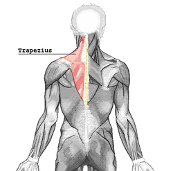
 muscles connecting the upper extremity to the vertebral column; trapezius is labeled at upper left. | |
| Details | |
| Origin | spinous processes of vertebrae C7-T12, Occipital Bone |
| Insertion | external occipital protuberance, nuchal ligament, medial superior nuchal line, posterior border of the lateral third of the clavicle, acromion process, and spine of scapula |
| Artery | superficial branch of transverse cervical artery or superficial cervical artery [1] |
| Nerve |
accessory nerve (motor) cervical spinal nerves C3 and C4 (motor and sensation)[2] |
| Actions | rotation, retraction, elevation, and depression of scapula |
| Antagonist | serratus anterior muscle, Latissimus dorsi |
| Identifiers | |
| Latin | musculus trapezius |
| TA | A04.3.01.001 |
| FMA | 9626 |
In human anatomy, the trapezius (/trəˈpiːzi.əs/) is one of two large superficial muscles that extend longitudinally from the occipital bone to the lower thoracic vertebrae and laterally to the spine of the scapula (shoulder blade). Its functions are to move the scapulae and support the arm.
The trapezius has three functional regions: the superior region (descending part), which supports the weight of the arm; the intermediate region (transverse part), which retracts the scapulae; and the inferior region (ascending part), which medially rotates and depresses the scapulae.
Structure
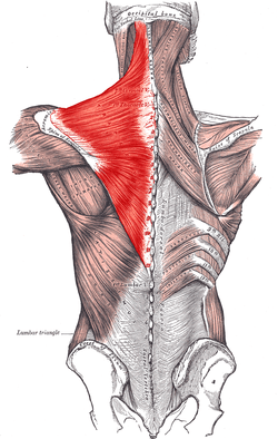
The trapezius muscle resembles a trapezium (trapezoid in American English), or diamond-shaped quadrilateral. The word "spinotrapezius" refers to the human trapezius, although it is not commonly used in modern texts. In other mammals, it refers to a portion of the analogous muscle.
The superior or upper (or descending) fibers of the trapezius originate from the spinous processes of C7, the external occipital protuberance, the medial third of the superior nuchal line of the occipital bone (both in the back of the head), and the ligamentum nuchae. From this origin they proceed downward and laterally to be inserted into the posterior border of the lateral third of the clavicle.
The middle fibers, or transverse of the trapezius arise from the spinous process of the seventh cervical (both in the back of the neck), and the spinous processes of the first, second, and third thoracic vertebrae. They are inserted into the medial margin of the acromion, and into the superior lip of the posterior border of the spine of the scapula.
The inferior or lower (or ascending) fibers of the trapezius arise from the spinous processes of the remaining thoracic vertebrae (T4–T12). From this origin they proceed upward and laterally to converge near the scapula and end in an aponeurosis, which glides over the smooth triangular surface on the medial end of the spine, to be inserted into a tubercle at the apex of this smooth triangular surface.
At its occipital origin, the trapezius is connected to the bone by a thin fibrous lamina, firmly adherent to the skin. The superficial and deep epimysia are continuous with an investing deep fascia that encircles the neck and also contains both sternocleidomastoid muscles.
At the middle, the muscle is connected to the spinous processes by a broad semi-elliptical aponeurosis, which reaches from the sixth cervical to the third thoracic vertebræ and forms, with that of the opposite muscle, a tendinous ellipse. The rest of the muscle arises by numerous short tendinous fibers.
Innervation
Motor function is supplied by the accessory nerve (CN XI). Sensation, including pain and proprioception, travel via the ventral rami of the third (C3) and fourth (C4) cervical nerves. Since it is a muscle of the upper limb, the trapezius is not innervated by dorsal rami despite being placed superficially in the back.
It is possible to feel the muscles of the superior trapezius become active by holding a weight in one hand in front of the body and, with the other hand, touching the area between the shoulder and the neck.
Function
Contraction of the trapezius muscle can have two effects: movement of the scapulae when the spinal origins are stable, and movement of the spine when the scapulae are stable. Its main function is to stabilize and move the scapula.
Scapular movements
The upper and lower fibres tend to rotate the scapula around the Sternoclavicular articulation so that the acromion and inferior angles move up and the medial border moves down. This rotation is in the opposite direction to that produced by levator scapulae and the rhomboids.
The middle fibres retract the scapula.
Spinal movements
When the scapulae are stable a co-contraction of both sides can extend the neck.
Exercises
- The upper portion of the trapezius can be developed by elevating the shoulders. Common exercises for this movement are any version of the clean, particularly the hang clean.
- Middle fibers are developed by pulling shoulder blades together. This adduction also uses the upper/lower fibers too.
- The lower part can be developed by drawing the shoulder blades downward while keeping the arms almost straight and stiff.
It is mainly used in throwing, with the deltoid muscles.
The upper and lower trapezius fibers also work in tandem with the serratus anterior to upwardly rotate the scapulae, such as during an overhead press. When activating together, the upper and lower fibers also assist the middle fibers (along with other muscles such as the rhomboids) with scapular retraction/adduction.
Muscle imbalances, which can heavily affect posture and compromise shoulder health, can result if all three sections of the trapezius not being developed equally.[3]
Many bodybuilders, including eight-time Mr. Olympia winner Ronnie Coleman, perform a maneuver known as a 'trap slap' (short for trapezius slap) before attempting to lift particularly heavy weights.[4] This technique involves a spotter slapping the lifter's upper back, with the desired effect of mentally preparing the recipient for their upcoming lift.[5] Variants of the trap slap include the 'lat slap' (short for latissimus dorsi slap), performed if the trapezius is inaccessible to the spotter (i.e. during a back squat).
Additional images
 Trapezius muscle.
 Trapezius muscle.
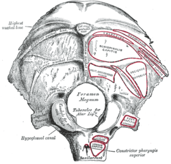
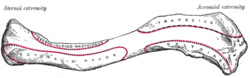 Left clavicle. Superior surface.
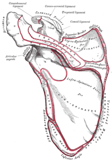 Left scapula. Posterior surface.
References
This article incorporates text in the public domain from the 20th edition of Gray's Anatomy (1918)
- ↑ "Tufts". Retrieved 2007-12-11.
- ↑ Arthur F. Dalley, Keith L. Moore, Anne M.R. Agur (2010). Clinically oriented anatomy (6th [International] ed.). Philadelphia [etc.]: Lippincott Williams & Wilkins, Wolters Kluwer. p. 700. ISBN 978-1-60547-652-0.
- ↑ Griffin, John C. (2006). Client-Centered Exercise Prescription. Champaign, IL: Human Kinetics. p. 217. ISBN 978-0-7360-5495-9.
- ↑ https://www.youtube.com/watch?v=LVVdlwf1iyM
- ↑ https://www.youtube.com/watch?v=4-Va6GdGHBw
External links
| Wikimedia Commons has media related to Trapezius muscle. |
- Muscles/TrapeziusUpper at exrx.net
- Trapezius (anatomy) at GPnotebook
- Superficial Back Dissection Video showing trapezius