CYB5R3
| View/Edit Human | |||
NADH-cytochrome b5 reductase 3 is an enzyme that in humans is encoded by the CYB5R3 gene.[2][3]
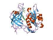
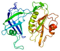
Structure
The CYB5R3 gene is located on the 22nd chromosome, with its specific location being 22q13.2. The gene contains 12 exons.[4] CYB5R3 encodes a 34.2 kDa protein that is composed of 301 amino acids; 63 peptides have been observed through mass spectrometry data.[5][6]
The entire gene is about 31 kb in length. Exon 2 contains the junction of the membrane-binding domain and the catalytic domain of b5R, which shows that there are two forms of b5R: a soluble form and a membrane-bound form. The 5' portion of this gene does not have typical regulatory transcriptional elements, but has the sequence G-G-G-C-G-G a total of five times. The GC content of this 5' portion of the gene is 86%, much higher than the average GC of the entire gene, which is 55%. There is also an atypical polyadenylation signal in the 3'-untranslated region of the gene.[7] The protein encoded by the CYB5R3 gene is cytochrome b5 reductase, a flavoprotein that is produced as two different isoforms with different localizations. There is an amphipathic microsomal isoform that is found in all cell types but red blood cells; this isoform has one hydrophobic membrane-anchoring domain and one catalytic domain that is hydrophilic. The other isoform, a soluble cytochrome b5 reductase isoform, is found in human erythrocytes. This protein is truncated, and encoded by an alternative transcript that produces only the larger, hydrophilic domain.[8] The protein contains 4 cyteine residues, Cys-203, -273, -283, and -297. Cys-283 is thought to be involved in NADH binding by chemical modification; in fact, both Cys-273 and Cys-283 are thought to be close to the NADH-binding site.[9] The NH2-terminal structure of the membrane-binding domain is CH3(CH2)12-CO-Gly-Ala-Gln-Leu-Ser-Thr-Leu-Gly-His-Met-Val-Leu-Phe-Pro-Val-Trp-Phe-Leu-Tyr-Ser-Leu-Leu-Met-Lys.[10]
Two forms of NADH-cytochrome b5 reductase are known, a membrane-bound form in somatic cells (anchored in the endoplasmic reticulum, mitochondria and other membranes) and a soluble form in erythrocytes. The membrane-bound form has both membrane-binding and catalytic domains. The soluble form has only the catalytic domain. This gene encodes both forms of the enzyme which arise from tissue-specific alternative transcripts that differ in the first exon. Mutations in this gene cause methemoglobinemias.[4]
Function
Cytochrome b5 reductase is involved in the transfer of reducing equivalents from the physiological electron donor, NADH, via an FAD domain to the small molecules of cytochrome b5. It’s also heavily involved in many oxidation and reduction reactions, such as the reduction of methemoglobin to hemoglobin.[8] Of the two forms of NADH-cytochrome b5 reductase, the membrane-bound form exists mainly on the cytoplasmic side of the endoplasmic reticulum and functions in desaturation and elongation of fatty acids, in cholesterol biosynthesis, and in drug metabolism. The erythrocyte form is located in a soluble fraction of circulating erythrocytes and is involved in methemoglobin reduction.[4]
Clinical significance
Mutations in the CYB5R3 gene cause methemoglobinemia types I and II. This is a rare autosomal recessive disease due to a deficiency of isoform of NADH-cytochrome b5 reductase.[11] Many mutations of this gene and the subsequent disease manifestation have been described.[12] The disease manifests as the accumulation of oxidized Fe+3 in humans.[8] Type I recessive congenital methemoglobinemia (RCM) is characterized by a deficiency of the soluble isoform and manifests as the cyanosis of skin and mucous membranes.[13] In type II, the defect affects both isoforms and thus affects more general tissues such as red blood cells, leukocytes, and all body tissues. This type is associated with mental deficiency and other neurologic symptoms, which may be because the cytochrome b5 system plays a crucial role in the desaturation of fatty acids in the body.[14] One patient was described as having a new class of this disorder, type III. This condition was characterized by a deficiency of NADH cytochrome b5 reductase in lymphocytes, platelets, and erythrocytes, but this was not associated with mental retardation.[15]
Interactions
CYB5R3 is known to interact with CYB5A, ENO1, and SUMO2 among other proteins.[4]
References
- ↑ "Human PubMed Reference:".
- ↑ Tomatsu S, Kobayashi Y, Fukumaki Y, Yubisui T, Orii T, Sakaki Y (Aug 1989). "The organization and the complete nucleotide sequence of the human NADH-cytochrome b5 reductase gene". Gene. 80 (2): 353–61. doi:10.1016/0378-1119(89)90299-0. PMID 2479590.
- ↑ Bull PC, Shephard EA, Povey S, Santisteban I, Phillips IR (Oct 1988). "Cloning and chromosomal mapping of human cytochrome b5 reductase (DIA1)". Annals of Human Genetics. 52 (Pt 4): 263–8. doi:10.1111/j.1469-1809.1988.tb01105.x. PMID 3268037.
- 1 2 3 4 "Entrez Gene: CYB5R3 cytochrome b5 reductase 3".
- ↑ ]Zong NC, Li H, Li H, Lam MP, Jimenez RC, Kim CS, Deng N, Kim AK, Choi JH, Zelaya I, Liem D, Meyer D, Odeberg J, Fang C, Lu HJ, Xu T, Weiss J, Duan H, Uhlen M, Yates JR, Apweiler R, Ge J, Hermjakob H, Ping P (Oct 2013). "Integration of cardiac proteome biology and medicine by a specialized knowledgebase". Circulation Research. 113 (9): 1043–53. doi:10.1161/CIRCRESAHA.113.301151. PMC 4076475
 . PMID 23965338.
 - ↑ "NADH-cytochrome b5 reductase 3". Cardiac Organellar Protein Atlas Knowledgebase (COPaKB).
- ↑ Tomatsu S, Kobayashi Y, Fukumaki Y, Yubisui T, Orii T, Sakaki Y (Aug 1989). "The organization and the complete nucleotide sequence of the human NADH-cytochrome b5 reductase gene". Gene. 80 (2): 353–61. doi:10.1016/0378-1119(89)90299-0. PMID 2479590.
- 1 2 3 Elahian F, Sepehrizadeh Z, Moghimi B, Mirzaei SA (Jun 2014). "Human cytochrome b5 reductase: structure, function, and potential applications". Critical Reviews in Biotechnology. 34 (2): 134–43. doi:10.3109/07388551.2012.732031. PMID 23113554.
- ↑ Shirabe K, Yubisui T, Nishino T, Takeshita M (Apr 1991). "Role of cysteine residues in human NADH-cytochrome b5 reductase studied by site-directed mutagenesis. Cys-273 and Cys-283 are located close to the NADH-binding site but are not catalytically essential". The Journal of Biological Chemistry. 266 (12): 7531–6. PMID 2019583.
- ↑ Murakami K, Yubisui T, Takeshita M, Miyata T (Feb 1989). "The NH2-terminal structures of human and rat liver microsomal NADH-cytochrome b5 reductases". Journal of Biochemistry. 105 (2): 312–7. PMID 2498303.
- ↑ Galeeva NM, Nenasheva SA, Kleĭmenova IS, Poliakov AV (Nov 2012). "[Novel large deletion c.22-1320_633+1224del in the CYB5R3 gene from patients with hereditary methemoglobinemia]". Genetika. 48 (11): 1336–46. PMID 23297489.
- ↑ Fermo E, Bianchi P, Vercellati C, Marcello AP, Garatti M, Marangoni O, Barcellini W, Zanella A (NaN). "Recessive hereditary methemoglobinemia: two novel mutations in the NADH-cytochrome b5 reductase gene". Blood Cells, Molecules & Diseases. 41 (1): 50–5. doi:10.1016/j.bcmd.2008.02.002. PMID 18343696. Check date values in:
|date=(help) - ↑ Galeeva NM, Voevoda MI, Spiridonova MG, Stepanov VA, Poliakov AV (Apr 2013). "[Population frequency and age of c.806C > T mutation in CYB5R3 gene as cause of recessive congenital methemoglobinemia in Yakutia]". Genetika. 49 (4): 523–30. PMID 23866629.
- ↑ Hudspeth MP, Joseph S, Holden KR (Jan 2010). "A novel mutation in type II methemoglobinemia". Journal of Child Neurology. 25 (1): 91–3. doi:10.1177/0883073809336136. PMID 19471045.
- ↑ Nagai T, Shirabe K, Yubisui T, Takeshita M (Feb 1993). "Analysis of mutant NADH-cytochrome b5 reductase: apparent "type III" methemoglobinemia can be explained as type I with an unstable reductase". Blood. 81 (3): 808–14. PMID 8427971.
Further reading
- Narahara K, Takahashi Y, Murakami M, Tsuji K, Yokoyama Y, Murakami R, Ninomiya S, Seino Y (Jun 1992). "Terminal 22q deletion associated with a partial deficiency of arylsulphatase A". Journal of Medical Genetics. 29 (6): 432–3. doi:10.1136/jmg.29.6.432. PMC 1016000. PMID 1352356.
- Dailey HA, Strittmatter P (Jun 1979). "Modification and identification of cytochrome b5 carboxyl groups involved in protein-protein interaction with cytochrome b5 reductase". The Journal of Biological Chemistry. 254 (12): 5388–96. PMID 221468.
- Malkinson AM, Siegel D, Forrest GL, Gazdar AF, Oie HK, Chan DC, Bunn PA, Mabry M, Dykes DJ, Harrison SD (Sep 1992). "Elevated DT-diaphorase activity and messenger RNA content in human non-small cell lung carcinoma: relationship to the response of lung tumor xenografts to mitomycin Cł". Cancer Research. 52 (17): 4752–7. PMID 1324793.
- Shirabe K, Yubisui T, Borgese N, Tang CY, Hultquist DE, Takeshita M (Oct 1992). "Enzymatic instability of NADH-cytochrome b5 reductase as a cause of hereditary methemoglobinemia type I (red cell type)". The Journal of Biological Chemistry. 267 (28): 20416–21. PMID 1400360.
- Katsube T, Sakamoto N, Kobayashi Y, Seki R, Hirano M, Tanishima K, Tomoda A, Takazakura E, Yubisui T, Takeshita M (Apr 1991). "Exonic point mutations in NADH-cytochrome B5 reductase genes of homozygotes for hereditary methemoglobinemia, types I and III: putative mechanisms of tissue-dependent enzyme deficiency". American Journal of Human Genetics. 48 (4): 799–808. PMC 1682939. PMID 1707593.
- Yubisui T, Shirabe K, Takeshita M, Kobayashi Y, Fukumaki Y, Sakaki Y, Takano T (Jan 1991). "Structural role of serine 127 in the NADH-binding site of human NADH-cytochrome b5 reductase". The Journal of Biological Chemistry. 266 (1): 66–70. PMID 1898726.
- Shirabe K, Yubisui T, Nishino T, Takeshita M (Apr 1991). "Role of cysteine residues in human NADH-cytochrome b5 reductase studied by site-directed mutagenesis. Cys-273 and Cys-283 are located close to the NADH-binding site but are not catalytically essential". The Journal of Biological Chemistry. 266 (12): 7531–6. PMID 2019583.
- Kobayashi Y, Fukumaki Y, Yubisui T, Inoue J, Sakaki Y (Apr 1990). "Serine-proline replacement at residue 127 of NADH-cytochrome b5 reductase causes hereditary methemoglobinemia, generalized type". Blood. 75 (7): 1408–13. PMID 2107882.
- Strittmatter P, Hackett CS, Korza G, Ozols J (Dec 1990). "Characterization of the covalent cross-links of the active sites of amidinated cytochrome b5 and NADH:cytochrome b5 reductase". The Journal of Biological Chemistry. 265 (35): 21709–13. PMID 2123873.
- Murakami K, Yubisui T, Takeshita M, Miyata T (Feb 1989). "The NH2-terminal structures of human and rat liver microsomal NADH-cytochrome b5 reductases". Journal of Biochemistry. 105 (2): 312–7. PMID 2498303.
- Yubisui T, Naitoh Y, Zenno S, Tamura M, Takeshita M, Sakaki Y (Jun 1987). "Molecular cloning of cDNAs of human liver and placenta NADH-cytochrome b5 reductase". Proceedings of the National Academy of Sciences of the United States of America. 84 (11): 3609–13. doi:10.1073/pnas.84.11.3609. PMC 304924. PMID 3035541.
- Yubisui T, Miyata T, Iwanaga S, Tamura M, Takeshita M (Feb 1986). "Complete amino acid sequence of NADH-cytochrome b5 reductase purified from human erythrocytes". Journal of Biochemistry. 99 (2): 407–22. PMID 3700359.
- Passon PG, Hultquist DE (Jul 1972). "Soluble cytochrome b 5 reductase from human erythrocytes". Biochimica et Biophysica Acta. 275 (1): 62–73. doi:10.1016/0005-2728(72)90024-2. PMID 4403130.
- Yubisui T, Miyata T, Iwanaga S, Tamura M, Yoshida S, Takeshita M, Nakajima H (Aug 1984). "Amino acid sequence of NADH-cytochrome b5 reductase of human erythrocytes". Journal of Biochemistry. 96 (2): 579–82. PMID 6389526.
- Shirabe K, Landi MT, Takeshita M, Uziel G, Fedrizzi E, Borgese N (Aug 1995). "A novel point mutation in a 3' splice site of the NADH-cytochrome b5 reductase gene results in immunologically undetectable enzyme and impaired NADH-dependent ascorbate regeneration in cultured fibroblasts of a patient with type II hereditary methemoglobinemia". American Journal of Human Genetics. 57 (2): 302–10. PMC 1801527. PMID 7668255.
- Vieira LM, Kaplan JC, Kahn A, Leroux A (Apr 1995). "Four new mutations in the NADH-cytochrome b5 reductase gene from patients with recessive congenital methemoglobinemia type II". Blood. 85 (8): 2254–62. PMID 7718898.
- Shirabe K, Fujimoto Y, Yubisui T, Takeshita M (Feb 1994). "An in-frame deletion of codon 298 of the NADH-cytochrome b5 reductase gene results in hereditary methemoglobinemia type II (generalized type). A functional implication for the role of the COOH-terminal region of the enzyme". The Journal of Biological Chemistry. 269 (8): 5952–7. PMID 8119939.
PDB gallery | ||||||
|---|---|---|---|---|---|---|
| ||||||