D-dimer
D-dimer (or D dimer) is a fibrin degradation product (or FDP), a small protein fragment present in the blood after a blood clot is degraded by fibrinolysis. It is so named because it contains two D fragments of the fibrin protein joined by a cross-link.[1]
D-dimer concentration may be determined by a blood test to help diagnose thrombosis. Since its introduction in the 1990s, it has become an important test performed in patients with suspected thrombotic disorders. While a negative result practically rules out thrombosis, a positive result can indicate thrombosis but does not rule out other potential causes. Its main use, therefore, is to exclude thromboembolic disease where the probability is low. In addition, it is used in the diagnosis of the blood disorder disseminated intravascular coagulation.[1]
Principles
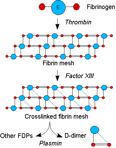
Coagulation, the formation of a blood clot or thrombus, occurs when the proteins of the coagulation cascade are activated, either by contact with damaged blood vessel wall and exposure to collagen in the tissue space (extrinsic pathway) or by activation of factor VII by tissue activating factors (intrinsic pathway). Both pathways lead to the generation of thrombin, an enzyme that turns the soluble blood protein fibrinogen into fibrin, which aggregates into proteofibrils. Another thrombin-generated enzyme, factor XIII, then crosslinks the fibrin proteofibrils at the D fragment site, leading to the formation of an insoluble gel which serves as a scaffold for blood clot formation.[1]
The circulating enzyme plasmin, the main enzyme of fibrinolysis, cleaves the fibrin gel in a number of places. The resultant fragments, "high molecular weight polymers", are digested several times more by plasmin to lead to intermediate and then to small polymers (fibrin degradation products or FDPs). The cross-link between two D fragments remains intact, however, and these are exposed on the surface when the fibrin fragments are sufficiently digested. The typical D-dimer containing fragment contains two D domains and one E domain of the original fibrinogen molecule.[1]
D-dimers are not normally present in human blood plasma, except when the coagulation system has been activated, for instance because of the presence of thrombosis or disseminated intravascular coagulation. The D-dimer assay depends on the binding of a monoclonal antibody to a particular epitope on the D-dimer fragment. Several detection kits are commercially available; all of them rely on a different monoclonal antibody against D-dimer. For some of these, the area of the D-dimer to which the antibody binds is known. The binding of the antibody is then measured quantitatively by one of various laboratory methods.[1]
Indications
D-dimer testing is of clinical use when there is a suspicion of deep venous thrombosis (DVT), pulmonary embolism (PE) or disseminated intravascular coagulation (DIC).[2] It is under investigation in the diagnosis of aortic dissection.[3][4]
For DVT and PE, there are possible various scoring systems that are used to determine the a priori clinical probability of these diseases; the best-known is the Wells score.[5]
- For a very high score, or pretest probability, a D-dimer will make little difference and anticoagulant therapy will be initiated regardless of test results, and additional testing for DVT or pulmonary embolism may be performed.
- For a moderate or low score, or pretest probability:
- A negative D-dimer test will virtually rule out thromboembolism: the degree to which the D-dimer reduces the probability of thrombotic disease is dependent on the test properties of the specific test used in the clinical setting: most available D-dimer tests with a negative result will reduce the probability of thromboembolic disease to less than 1% if the pretest probability is less than 15-20%. Chest computed tomography (CT angiography) should not be used to evaluate pulmonary embolism for persons with negative results of a D-dimer assay.[6]
- If the D-dimer reads high, then further testing (ultrasound of the leg veins or lung scintigraphy or CT scanning) is required to confirm the presence of thrombus. Anticoagulant therapy may be started at this point or withheld until further tests confirm the diagnosis, depending on the clinical situation.
In some hospitals, they are measured by laboratories after a form is completed showing the probability score and only if the probability score is low or intermediate. This reduces the need for unnecessary tests in those who are high-probability.[7] Performing the D-dimer test first can avoid a significant proportion of imaging tests and is less invasive. Since the D-dimer can exclude the need for imaging, specialty professional organizations recommend that physicians use D-dimer testing as an initial diagnostic.[8][9][10][11]
Interpretation
Reference ranges
Following are reference ranges for D-dimer:[12]
| Units | Nonpregnant adult | First trimester | Second trimester | Third trimester |
|---|---|---|---|---|
| mg/L or µg/mL | < 0.5 | 0.05 - 0.95 | 0.32 - 1.29 | 0.13 -1.7 |
| µg/L or ng/mL | < 500 | 50 - 950 | 320 - 1290 | 130 - 1700 |
| nmol/L | < 2.7 | 0.3 - 5.2 | 1.8 - 7.1 | 0.7 - 9.3 |
Thrombotic disease
Various kits have a 93-95% sensitivity (true positive rate). For hospitalized patients, one study found the specificity to be about 50% (true negative rate) in the diagnosis of thrombotic disease.[13]
- False positive readings can be due to various causes: liver disease, high rheumatoid factor, inflammation, malignancy, trauma, pregnancy, recent surgery as well as advanced age.[14]
- False negative readings can occur if the sample is taken either too early after thrombus formation or if testing is delayed for several days. Additionally, the presence of anti-coagulation can render the test negative because it prevents thrombus extension. The anti-coagulation medications dabigatran and rivaroxaban decrease D-dimer levels but do not interfere with the D-dimer assay.[15]
- False values may be obtained if the specimen collection tube is not sufficiently filled (false low value if underfilled and false high value if overfilled). This is due to the dilutional effect of the anticoagulant (the blood must be collected in a 9:1 blood to anticoagulant ratio).
- Likelihood ratios are derived from sensitivity and specificity to adjust pretest probability.
In interpretation of the d-dimer, for patients over age 50 a value of (patient's age) x 10 μg/l may be abnormal.[16][17]
History
D-dimer was originally described in the 1970s, and found its diagnostic application in the 1990s.[1]
References
- 1 2 3 4 5 6 7 Adam SS, Key NS, Greenberg CS (March 2009). "D-dimer antigen: current concepts and future prospects". Blood. 113 (13): 2878–2887. doi:10.1182/blood-2008-06-165845. PMID 19008457.
- ↑ General Practice Notebook > D-dimer Retrieved September 2011
- ↑ Suzuki T, Distante A, Eagle K (2010). "Biomarker-assisted diagnosis of acute aortic dissection: How far we have come and what to expect". Current Opinion in Cardiology. 25 (6): 541–545. doi:10.1097/HCO.0b013e32833e6e13. PMID 20717014.
- ↑ Ranasinghe AM, Bonser RS (2010). "Biomarkers in Acute Aortic Dissection and Other Aortic Syndromes". Journal of the American College of Cardiology. 56 (19): 1535–1541. doi:10.1016/j.jacc.2010.01.076. PMID 21029872.
- ↑ Wells PS, Anderson DR, Rodger M, Forgie M, Kearon C, Dreyer J, Kovacs G, Mitchell M, Lewandowski B, Kovacs MJ (2003). "Evaluation of D-dimer in the diagnosis of suspected deep-vein thrombosis". N. Engl. J. Med. 349 (13): 1227–1235. doi:10.1056/NEJMoa023153. PMID 14507948.
- ↑ -->American College of Chest Physicians; American Thoracic Society (September 2013), "Five Things Physicians and Patients Should Question", Choosing Wisely: an initiative of the ABIM Foundation, American College of Chest Physicians and American Thoracic Society, retrieved 6 January 2013.
- ↑ Rathbun SW, Whitsett TL, Vesely SK, Raskob GE (2004). "Clinical utility of D-dimer in patients with suspected pulmonary embolism and nondiagnostic lung scans or negative CT findings". Chest. 125 (3): 851–855. doi:10.1378/chest.125.3.851. PMC 1215466
 . PMID 15006941.
 - ↑ American College of Physicians, presented by ABIM Foundation, "Five Things Physicians and Patients Should Question" (PDF), Choosing Wisely, American College of Physicians, retrieved August 14, 2012
- ↑ Fesmire FM, Brown MD, Espinosa JA, Shih RD, Silvers SM, Wolf SJ, Decker WW (2011). "Critical Issues in the Evaluation and Management of Adult Patients Presenting to the Emergency Department with Suspected Pulmonary Embolism". Annals of Emergency Medicine. 57 (6): 628–652.e75. doi:10.1016/j.annemergmed.2011.01.020. PMID 21621092.
- ↑ Torbicki A, Perrier A, Konstantinides S, Agnelli G, Galiè N, Pruszczyk P, Bengel F, Brady AJ, Ferreira D, Janssens U, Klepetko W, Mayer E, Remy-Jardin M, Bassand JP (2008). "Guidelines on the diagnosis and management of acute pulmonary embolism: The Task Force for the Diagnosis and Management of Acute Pulmonary Embolism of the European Society of Cardiology (ESC)". European Heart Journal. 29 (18): 2276–2315. doi:10.1093/eurheartj/ehn310. PMID 18757870.
- ↑ Qaseem A, Snow V, Barry P, Hornbake ER, Rodnick JE, Tobolic T, Ireland B, Segal J, Bass E, Weiss KB, Green L, Owens DK (2007). "Current Diagnosis of Venous Thromboembolism in Primary Care: A Clinical Practice Guideline from the American Academy of Family Physicians and the American College of Physicians". The Annals of Family Medicine. 5 (1): 57–62. doi:10.1370/afm.667. PMID 17261865.
- ↑ Reference Values During Pregnancy at perinatology.com. Retrieved October 2014.
- ↑ Schrecengost JE, LeGallo RD, Boyd JC, Moons KG, Gonias SL, Rose CE, Bruns DE (September 2003). "Comparison of diagnostic accuracies in outpatients and hospitalized patients of D-dimer testing for the evaluation of suspected pulmonary embolism". Clinical Chemistry. 49 (9): 1483–1490. doi:10.1373/49.9.1483. PMID 12928229.
- ↑ Kabrhel C, Mark Courtney D, Camargo CA, Plewa MC, Nordenholz KE, Moore CL, Richman PB, Smithline HA, Beam DM, Kline JA (14 May 2010). "Factors Associated With Positive D-dimer Results in Patients Evaluated for Pulmonary Embolism". Academic Emergency Medicine. 17 (6): 589–597. doi:10.1111/j.1553-2712.2010.00765.x. PMC 3538031. PMID 20624138.
- ↑ Baglin, Trevor; Keeling, David; Kitchen, Steve (2012). "Effects on routine coagulation screens and assessment of anticoagulant intensity in patients taking oral dabigatran or rivaroxaban: Guidance from the British Committee for Standards in Haematology". British Journal of Haematology. 159 (4): 427–429. doi:10.1111/bjh.12052. ISSN 0007-1048.
- ↑ van Es J, Mos I, Douma R, Erkens P, Durian M, Nizet T, van Houten A, Hofstee H, ten Cate H, Ullmann E, Büller H, Huisman M, Kamphuisen PW (2012). "The combination of four different clinical decision rules and an age-adjusted D-dimer cut-off increases the number of patients in whom acute pulmonary embolism can safely be excluded.". Thromb Haemost. 107 (1): 167–71. doi:10.1160/TH11-08-0587. PMID 22072293.
- ↑ Douma RA, le Gal G, Söhne M, Righini M, Kamphuisen PW, Perrier A, Kruip MJ, Bounameaux H, Büller HR, Roy PM (2010). "Potential of an age adjusted D-dimer cut-off value to improve the exclusion of pulmonary embolism in older patients: a retrospective analysis of three large cohorts.". BMJ. 340: c1475. doi:10.1136/bmj.c1475. PMC 2847688. PMID 20354012.
External links
- D-dimer - Lab Tests Online