Dark field microscopy
Dark field microscopy (dark ground microscopy) describes microscopy methods, in both light and electron microscopy, which exclude the unscattered beam from the image. As a result, the field around the specimen (i.e., where there is no specimen to scatter the beam) is generally dark.

Light microscopy applications
In optical microscopy, darkfield describes an illumination technique used to enhance the contrast in unstained samples. It works by illuminating the sample with light that will not be collected by the objective lens, and thus will not form part of the image. This produces the classic appearance of a dark, almost black, background with bright objects on it.
The light's path
The steps are illustrated in the figure where an inverted microscope is used.
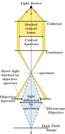
- Light enters the microscope for illumination of the sample.
- A specially sized disc, the patch stop (see figure) blocks some light from the light source, leaving an outer ring of illumination. A wide phase annulus can also be reasonably substituted at low magnification.
- The condenser lens focuses the light towards the sample.
- The light enters the sample. Most is directly transmitted, while some is scattered from the sample.
- The scattered light enters the objective lens, while the directly transmitted light simply misses the lens and is not collected due to a direct illumination block (see figure).
- Only the scattered light goes on to produce the image, while the directly transmitted light is omitted.
Advantages and disadvantages

Dark field microscopy is a very simple yet effective technique and well suited for uses involving live and unstained biological samples, such as a smear from a tissue culture or individual, water-borne, single-celled organisms. Considering the simplicity of the setup, the quality of images obtained from this technique is impressive.
The main limitation of dark field microscopy is the low light levels seen in the final image. This means the sample must be very strongly illuminated, which can cause damage to the sample. Dark field microscopy techniques are almost entirely free of artifacts, due to the nature of the process. However, the interpretation of dark field images must be done with great care, as common dark features of bright field microscopy images may be invisible, and vice versa.
While the dark field image may first appear to be a negative of the bright field image, different effects are visible in each. In bright field microscopy, features are visible where either a shadow is cast on the surface by the incident light, or a part of the surface is less reflective, possibly by the presence of pits or scratches. Raised features that are too smooth to cast shadows will not appear in bright field images, but the light that reflects off the sides of the feature will be visible in the dark field images.
- Comparison of transillumination techniques used to generate contrast in a sample of tissue paper. 1.559 μm/pixel (when viewed at full resolution).
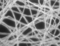 Dark field illumination, sample contrast comes from light scattered by the sample.
 Bright field illumination, sample contrast comes from absorbance of light in the sample.
 Cross-polarized light illumination, sample contrast comes from rotation of polarized light through the sample.
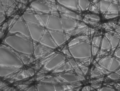 Phase contrast illumination, sample contrast comes from interference of different path lengths of light through the sample.
Use in computing
Dark field microscopy has recently been used in computer mouse pointing devices, in order to allow an optical mouse to work on transparent glass by imaging microscopic flaws and dust on its surface.
Dark field microscopy combined with hyperspectral imaging
When coupled to hyperspectral modality, dark field microscopy becomes a powerful tool for the characterization of nanomaterials embedded in cells. In a recent publication, Patskovsky et al. used this technique to study the attachment of gold nanoparticles (AuNPs) targeting CD44+ cancer cells. [1]
Transmission electron microscope applications
Darkfield studies in transmission electron microscopy play a powerful role in the study of crystals and crystal defects, as well as in the imaging of individual atoms.
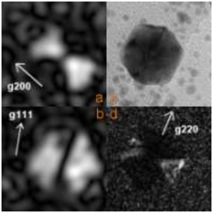
Conventional darkfield imaging
Briefly, imaging[2] involves tilting the incident illumination until a diffracted, rather than the incident, beam passes through a small objective aperture in the objective lens back focal plane. Darkfield images, under these conditions, allow one to map the diffracted intensity coming from a single collection of diffracting planes as a function of projected position on the specimen, and as a function of specimen tilt.
In single crystal specimens, single-reflection darkfield images of a specimen tilted just off the Bragg condition allow one to "light up" only those lattice defects, like dislocations or precipitates, which bend a single set of lattice planes in their neighborhood. Analysis of intensities in such images may then be used to estimate the amount of that bending. In polycrystalline specimens, on the other hand, darkfield images serve to light up only that subset of crystals which is Bragg reflecting at a given orientation.
Animation: darkfield imaging of crystals  Digital darkfield simulation of 2nm metal particles on a nano-cylinder
Weak beam imaging
Weak beam imaging involves optics similar to conventional darkfield, but use of a diffracted beam harmonic rather than the diffracted beam itself. Much higher resolution of strained regions around defects can be obtained in this way.
Low and high angle annular darkfield imaging
Annular dark-field imaging requires one to form images with electrons diffracted into an annular aperture centered on, but not including, the unscattered beam. For large scattering angles in a scanning transmission electron microscope, this is sometimes called Z-contrast imaging because of the enhanced scattering from high atomic number atoms.
Digital darkfield analysis
This a mathematical technique intermediate between direct and reciprocal (Fourier-transform) space for exploring images with well-defined periodicities, like electron microscope lattice-fringe images. As with analog darkfield imaging in a transmission electron microscope, it allows one to "light up" those objects in the field of view where periodicities of interest reside. Unlike analog darkfield imaging it may also allow one to map the Fourier-phase of periodicities, and hence phase-gradients which provide quantitative information on vector lattice-strain.
See also
Footnotes
- ↑ S. Patskovsky; et al. (2014). "Wide-field hyperspectral 3D imaging of functionalized gold nanoparticles targeting cancer cells by reflected light microscopy". Biophotonics: 1–7. doi:10.1002/jbio.201400025.
- ↑ P. Hirsch, A. Howie, R. Nicholson, D. W. Pashley and M. J. Whelan (1965/1977) Electron microscopy of thin crystals (Butterworths/Krieger, London/Malabar FL) ISBN 0-88275-376-2
External links
| Library resources about Dark field microscopy |
| Wikimedia Commons has media related to Dark-field microscopy. |
- Nikon - Stereomicroscopy > Darkfield Illumination
- Molecular Expressions
- Darkfield Illumination Primer
- Gage SH. 1920. Modern dark-field microscopy and the history of its development. Transactions of the American Microscopical Society 39(2):95–141.
- animations and explanations on various types of microscopes including dark field microscopes (Université Paris Sud)
Optical microscopy | ||
|---|---|---|
| Illumination and contrast methods |  | |
| Fluorescence methods | ||
| Sub-diffraction limit techniques | ||