Foramen lacerum
| Foramen lacerum | |
|---|---|
 | |
| Details | |
| Identifiers | |
| Latin | Foramen lacerum |
| TA | A02.1.00.055 |
| FMA | 54809 |
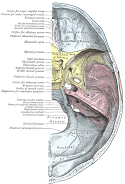
The foramen lacerum (Latin for lacerated piercing) is a triangular hole in the base of the skull located between the sphenoid, apex of petrous temporal and basilar part of occipital.
Structure
The foramen lacerum is a foramen situated anteromedial to the carotid canal.[1] :776
Development
The foramen lacerum fills with cartilage after birth.[1] :776
Function
The artery of pterygoid canal, the nerve of pterygoid canal and some venous drainage pass through the foramen lacerum.
- In the foramen lacerum the greater petrosal nerve joins with the deep petrosal nerve to form the nerve of the pterygoid canal. The deep petrosal nerve carries sympathetic and the greater petrosal nerve carries parasympathetic fibers of the autonomic nervous system to blood vessels, mucous membranes, salivary glands, and lacrimal glands.
- Furthermore, one of the terminal branches of the ascending pharyngeal artery (itself a branch of the external carotid artery) passes through the foramen lacerum. The ascending pharyngeal artery is one of three possible "meningeal branches" of this vessel.
- Some emissary veins pass through the foramen lacerum. These connect the extracranial pterygoid plexus with the intracranial cavernous sinus and present an unopposed route for infection.
The internal carotid artery passes from the carotid canal in the base of the skull, emerging and coursing superior to foramen lacerum as it exits the carotid canal. The internal carotid artery does not travel through foramen lacerum. The segment of the internal carotid artery that travels above foramen lacerum is called the lacerum segment.
Clinical relevance
The Foramen lacerum is a potential route for nasopharyngeal carcinoma to gain access to the cavernous sinus and affect cranial nerves.[2]
Additional images
-
Foramen lacerum
References
- 1 2 Drake, Richard L.; Vogl, Wayne; Tibbitts, Adam W.M. Mitchell; illustrations by Richard; Richardson, Paul (2005). Gray's anatomy for students. Philadelphia: Elsevier/Churchill Livingstone. ISBN 978-0-8089-2306-0.
- ↑ Christodouleas, Boris Hristov, Steven H. Lin, John P. (2010). Radiation oncology : a question-based review. Philadelphia, Pa.: Lippincott Williams & Wilkins. p. 138. ISBN 1608314448.
External links
- Anatomy figure: 22:5b-10 at Human Anatomy Online, SUNY Downstate Medical Center - "Internal view of skull."
- Photo of model at Waynesburg College skeleton/foramenlacerum
- cranialnerves at The Anatomy Lesson by Wesley Norman (Georgetown University) (VII)
- Tauber M, van Loveren H, Jallo G, Romano A, Keller J (1999). "The enigmatic foramen lacerum". Neurosurgery. 44 (2): 386–91; discussion 391–3. doi:10.1097/00006123-199902000-00083. PMID 9932893.
- Anatomy diagram: 34257.000-1 at Roche Lexicon - illustrated navigator, Elsevier
- Image at ucsd.edu