Facial canal
| Facial canal | |
|---|---|
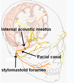
 Route of facial nerve, with facial canal labeled | |
|
View of the inner wall of the tympanum. (Facial canal visible in upper left.) | |
| Details | |
| Identifiers | |
| Latin | canalis nervi facialis, canalis facialis |
| TA | A02.1.06.009 |
| FMA | 54952 |
The facial canal (Canalis nervi facialis)(also known as the Fallopian Canal[1] - first described by Gabriele Falloppio-) is a Z-shaped canal running through the temporal bone from the internal acoustic meatus to the stylomastoid foramen. In humans it is approximately 3 centimeters long, which makes it the longest human osseous canal of a nerve.[2] It is located within the middle ear region, according to its shape it is divided into three main segments: the labyrinthine, the tympanic, and the mastoidal segment.[3] It contains Cranial Nerve VII, also known as the facial nerve.
See also
Additional Images
-
Lateral head anatomy detail. Facial nerve dissection.
-

Tympanic cavity. Facial canal. Internal carotid artery.
References
- ↑ Abing W, Rauchfuss A (2005). "Fetal development of the tympanic part of the facial canal". European Archives of Oto-Rhino-Laryngology. 243 (6): 374–377. PMID 3566620.
- ↑ Weiglein AH (June 1996). "Postnatal development of the facial canal. An investigation based on cadaver dissections and computed tomography". Surgical and Radiologic Anatomy. 18 (2): 115–23. doi:10.1007/BF01795229. PMID 8782317.
- ↑ Weiglein AH, Anderhuber W, Jakse R, Einspieler R (1994). "Imaging of the facial canal by means of multiplanar angulated 2-D-high-resolution CT-reconstruction". Surgical and radiologic anatomy. 16 (4): 423–427. doi:10.1007/BF01627665. PMID 7725200.
External links
This article is issued from Wikipedia - version of the 8/7/2016. The text is available under the Creative Commons Attribution/Share Alike but additional terms may apply for the media files.