Haversian canal
| Haversian canal | |
|---|---|
|
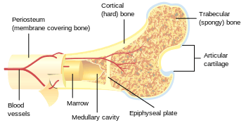
Diagram of compact bone from a transverse section of a typical long bone's cortex. | |

Haversian canals[lower-roman 1] (sometimes canals of Havers, named after British physician Clopton Havers) are a series of microscopic tubes in the outermost region of bone called cortical bone that allow blood vessels and nerves to travel through them. Each haversian canal generally contains one or two capillaries and nerve fibres. The channels are formed by concentric layers called lamellae. The haversian canals surround blood vessels and nerve cells throughout bones and communicate with bone cells (contained in spaces within the dense bone matrix called lacunae) through connections called canaliculi. This unique arrangement is conducive to mineral salt deposits and storage which gives bone tissue its strength.
In mature compact bone most of the individual lamellae form concentric rings around larger longitudinal canals (approx. 50 µm in diameter) within the bone tissue. These canals are called haversian canals. Haversian canals are contained within osteons, which are typically arranged along the long axis of the bone in parallel to the surface. The canals and the surrounding lamellae (8-15) form the functional unit, called a haversian system or osteon.
Notes
- ↑ As with other medical eponyms, the adjective derived from the eponym's name is usually lowercased; thus haversian (but canal of Havers), fallopian, eustachian, and parkinsonism (but Parkinson disease); for more, see eponym > orthographic conventions.
References
External links
- http://www.lab.anhb.uwa.edu.au/mb140/
- Youtube, YouTube video of Haversian canal system within cortical bone
Additional images
- Bone by decalcification (40x):
- Volkmann's canal
- Haversian canal
- Blood vessel
- Bone by decalcification (100x):
- Volkmann's canal
- Haversian canal
- lamella
- Lacuna
