Humerus
| Humerus | |
|---|---|
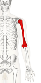
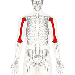 Position of humerus (shown in red) from an anterior viewpoint | |
| Details | |
| Identifiers | |
| Latin | humerus |
| MeSH | A02.835.232.087.090.400 |
| TA | A02.4.04.001 |
| FMA | 13303 |
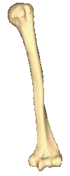
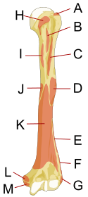
The humerus (/ˈhjuːmərəs/, Plural: humeri) is a long bone in the arm or forelimb that runs from the shoulder to the elbow. It connects the scapula and the two bones of the lower arm, the radius and ulna, and consists of three sections. The humeral upper extremity consists of a rounded head, a narrow neck, and two short processes (tubercles, sometimes called tuberosities). The body is cylindrical in its upper portion, and more prismatic below. The lower extremity consists of 2 epicondyles, 2 processes (trochlea & capitulum), and 3 fossae (radial fossa, coronoid fossa, and olecranon fossa). As well as its true anatomical neck, the constriction below the greater and lesser tubercles of the humerus is referred to as its surgical neck due to its tendency to fracture, thus often becoming the focus of surgeons.
Etymology
The word "humerus" is derived from Latin: humerus, umerus meaning upper arm, shoulder, and is linguistically related to Gothic ams shoulder and Greek ōmos.[1]
Structure
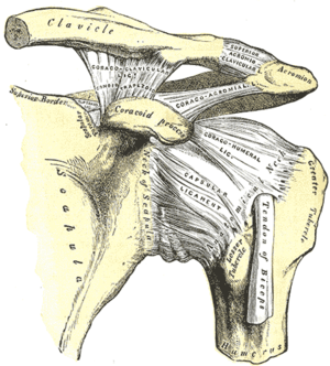
Articulations
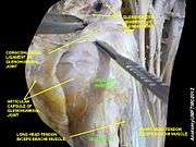
At the shoulder, the head of the humerus articulates with the glenoid fossa of the scapula. More distally, at the elbow, the capitulum of the humerus articulates with the head of the radius, and the trochlea of the humerus articulates with the trochlear notch of the ulna.
|


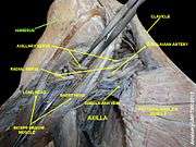
Nerves
The axillary nerve is located at the proximal end, against the shoulder girdle. Dislocation of the humerus's glenohumeral joint, has the potential to injure the axillary nerve or the axillary artery. Signs and symptoms of this dislocation include a loss of the normal shoulder contour and a palpable depression under the acromion.
The radial nerve follows the humerus closely. At the midshaft of the humerus, the radial nerve travels from the posterior to the anterior aspect of the bone in the spiral groove. A fracture of the humerus in this region can result in radial nerve injury.
The ulnar nerve at the distal end of the humerus near the elbow is sometimes referred to in popular culture as 'the funny bone'. Striking this nerve can cause a tingling sensation ("funny" feeling), and sometimes a significant amount of pain. It lies posterior to the medial epicondyle, and is easily damaged in elbow injuries.[2]
|


Function
Muscular attachment
The deltoid originates on the lateral third of the clavicle, acromion and the crest of the spine of the scapula. It is inserted on the deltoid tuberosity of the humerus and has several actions including abduction, extension, and circumduction of the shoulder. The supraspinatus also originates on the spine of the scapula. It inserts on the greater tubercle of the humerus, and assists in abduction of the shoulder.
The pectoralis major, teres major, and latissimus dorsi insert at the intertubercular groove of the humerus. They work to adduct and medially, or internally, rotate the humerus.
The infraspinatus and teres minor insert on the greater tubercle, and work to laterally, or externally, rotate the humerus. In contrast, the subscapularis muscle inserts onto the lesser tubercle and works to medially, or internally, rotate the humerus.
The biceps brachii, brachialis, and brachioradialis (which attaches distally) act to flex the elbow. (The biceps do not attach to the humerus.) The triceps brachii and anconeus extend the elbow, and attach to the posterior side of the humerus.
The four muscles of supraspinatus, infraspinatus, teres minor and subscapularis form a musculo-ligamentous girdle called the rotator cuff. This cuff stabilizes the very mobile but inherently unstable glenohumeral joint. The other muscles are used as counterbalances for the actions of lifting/pulling and pressing/pushing.
.png)

.png) | 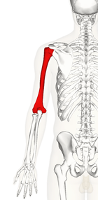 |  |  |  |
Other animals
Primitive fossils of amphibians had little, if any, shaft connecting the upper and lower extremities, making their limbs very short. In most living tetrapods, however, the humerus has a similar form to that of humans; connecting their extremities. In many reptiles and some primitive mammals, the lower extremity includes a large foramen, or opening, which allows nerves and blood vessels pass through.[3]
Additional images
 Position of humerus (shown in red). Animation.
 Left humerus. Animation.
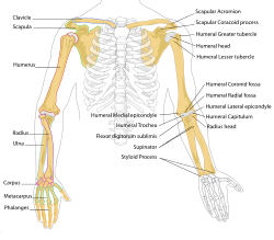 Human arm bones diagram.
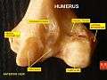 Humerus - inferior epiphysis. Anterior view.
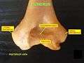 Trochlea. Posterior view.
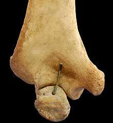 Humerus - inferior epiphysis. Posterior view.
 Humerus - superior epiphysis. Anterior view.
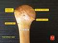 Humerus - superior epiphysis. Posterior view.
.jpg) Elbow joint. Deep dissection. Anterior view.
Elbow joint. Deep dissection. Anterior view. Elbow joint. Deep dissection. Posterior view.
 Elbow joint. Deep dissection. Posterior view.
See also
References
- This article incorporates text in the public domain from the 20th edition of Gray's Anatomy (1918)
External links
| Wikimedia Commons has media related to Humerus. |
-
 "Humerus". New International Encyclopedia. 1905.