Jones fracture
| Jones fracture | |
|---|---|
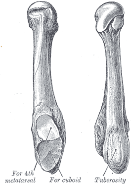 | |
| The fifth metatarsal. | |
| Classification and external resources | |
| Specialty | emergency medicine |
| ICD-10 | S92.3 |
| ICD-9-CM | 825.25, 825.35 |
| eMedicine | radio/850 |

A Jones fracture is a fracture in the meta-diaphyseal junction of the fifth metatarsal of the foot.[1] The proximal end of the metatarsal, where the Jones fracture occurs, is near the midportion of the foot, on the fifth ray (of which the 5th toe belongs). Those who sustain a Jones fracture have pain over this area, swelling, and difficulty walking. The fracture was first described by orthopedic surgeon Sir Robert Jones who sustained this injury himself (while dancing) and reported it in the Annals of Surgery in 1902.[2]
Diagnosis
A patient with a Jones fracture may not realize that a fracture has occurred. Diagnosis includes the palpation of an intact peroneus brevis tendon, and demonstration of local tenderness distal to the tuberosity of the fifth metatarsal, and localized over the diaphysis of the proximal metatarsal. Bony crepitus is unusual.
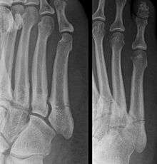
This injury should be differentiated from the developmental apophysis (5th metatarsal tuberosity) commonly and normally occurring at this site in adolescents. Differentiation is possible by characteristics such as absence of sclerosis of the fractured edges (in acute cases) and orientation of the lucent line: transverse (at 90 degrees) to the metatarsal axis for the fracture (due to avulsion pull by the peroneus brevis muscle inserting at the proximal tip) - and parallel to the metatarsal axis in the case of the apophysis. Diagnostic x-rays include anteroposterior, oblique, and lateral views and should be made with the foot in full flexion.
Treatment
A legitimate concern in any fracture is whether the fracture will heal quickly and without complication. Failure of the fractured ends to unite is called non-union and its frequency varies with the fracture site, some fracture sites being notorious for non-union. An example of such would be a scaphoid (navicular) fracture of the wrist. Such a complication also involves fractures of the proximal end of the fifth metatarsal, such as the Jones fracture. This has been the subject of interest, and initially led to the description of three zones at the proximal end of the fifth metatarsal.
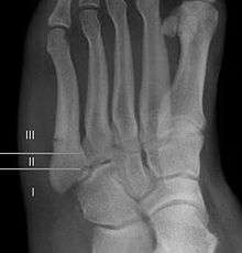
Zones I and II have been associated with relatively guaranteed union and this union has taken place with only limited restriction of activity combined with early immobilization. On the other hand, zone III has been associated with either delayed or non-union and, consequently, it has been generally agreed that fractures in this area should be considered for some form of internal immobilization, such as internal screw fixation. More recently, because of the similar behavior of the original zones I and II, it has been suggested that zones I and II be combined leading to current recommendations for two zones, zone I being associated with uncomplicated union, and zone II being prone to nonunion and therefore considered for internal fixation.

These zones can be identified anatomically and on x-ray adding to the clinical usefulness of this classification.[3] It should be emphasized that surgical intervention is not, by itself, a guarantee of cure and has its own complication rate. Other reviews of the literature have concluded that conservative, non-operative, treatment is an acceptable option for the non-athlete.[4]
If non-surgical management is pursued, a cast, splint or walking boot for four to eight weeks may be used. Three-fourths of fractures so treated will unite.
Prognosis
For several reasons, a Jones fracture may not unite. The diaphyseal bone (zone II), where the fracture occurs, is an area of potentially poor blood supply, existing in a watershed area between two blood supplies. This may compromise healing. In addition, there are various tendons, including the peroneus brevis and fibularis tertius, and two small muscles attached to the bone. These may pull the fracture apart and prevent healing.
Other proximal fifth metatarsal fractures
Other proximal fifth metatarsal fractures exist, although they are not as severe as a Jones fracture. If the fracture enters the intermetatarsal joint, it is a Jones fracture. If, however, it enters the tarsometatarsal joint, then it is an avulsion fracture caused by pull from the peroneus brevis. An avulsion fracture is sometimes called a Pseudo-Jones fracture or a Dancer's fracture.
Notes
- ↑ Joel A. DeLisa; Bruce M. Gans; Nicholas E. Walsh (2005). Physical Medicine and Rehabilitation: Principles and Practice. Lippincott Williams & Wilkins. pp. 881–. ISBN 978-0-7817-4130-9.
- ↑ Jones, Robert (Jun 1902). "I. Fracture of the Base of the Fifth Metatarsal Bone by Indirect Violence.". Ann Surg. 35 (6): 697–700. PMC 1425723
 . PMID 17861128.
 - ↑ Polzer H, Polzer S, Mutschler W, Prall WC (Oct 2012). "Acute fractures to the proximal fifth metatarsal bone: development of classification and treatment recommendations based on the current evidence". Injury. 43: 1626–32. doi:10.1016/j.injury.2012.03.010. PMID 22465516.
- ↑ Dean BJ, Kothari A, Uppal H, Kankate R (Aug 2012). "The jones fracture classification, management, outcome, and complications: a systematic review". Foot Ankle Spec. 5(4): 256–9. doi:10.1177/1938640012444730. PMID 22547534.
External links
| Wikimedia Commons has media related to X-rays of Jones fractures. |
- Jones fracture - wheelessonline.com