Lumbar vertebrae
| Lumbar vertebrae | |
|---|---|
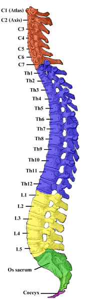
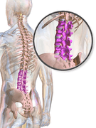
 Position of human lumbar vertebrae (shown in red). It consists of 5 bones, from top to down, L1, L2, L3, L4 and L5. | |
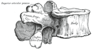
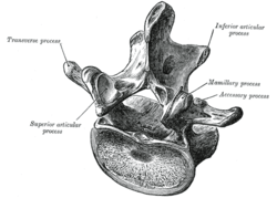 A typical lumbar vertebra | |
| Details | |
| Identifiers | |
| Latin | vertebrae lumbales |
| TA | A02.2.04.001 |
| FMA | 72065 |
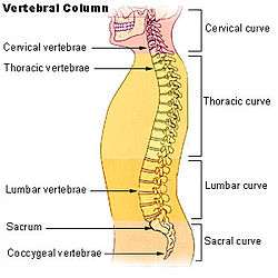
The lumbar vertebrae are, in human anatomy, the five vertebrae between the rib cage and the pelvis. They are the largest segments of the vertebral column and are characterized by the absence of the foramen transversarium within the transverse process (as it is only found in the cervical region), and by the absence of facets on the sides of the body (as only found in the thoracic region). They are designated L1 to L5, starting at the top. The lumbar vertebrae help support the weight of the body, and permit movement.
Human anatomy
General characteristics

The figure on the left depicts the general characteristics of the first through fourth lumbar vertebrae. The fifth vertebra contains certain peculiarities, which are detailed below.
As with other vertebrae, each lumbar vertebra consists of a vertebral body and a vertebral arch. The vertebral arch, consisting of a pair of pedicles and a pair of laminae, encloses the vertebral foramen (opening) and supports seven processes.
Body
The vertebral body of each lumbar vertebra is large, wider from side to side than from front to back, and a little thicker in front than in back. It is flattened or slightly concave above and below, concave behind, and deeply constricted in front and at the sides.[1]
Arch
The pedicles are very strong, directed backward from the upper part of the vertebral body; consequently, the inferior vertebral notches are of considerable depth.[1] The pedicles change in morphology from the upper lumbar to the lower lumbar. They increase in sagittal width from 9 mm to up to 18 mm at L5. They increase in angulation in the axial plane from 10 degrees to 20 degrees by L5. The pedicle is sometimes used as a portal of entrance into the vertebral body for fixation with pedicle screws or for placement of bone cement as with kyphoplasty or vertebroplasty.
The laminae are broad, short, and strong.[1] They form the posterior portion of the vertebral arch. In the upper lumbar region the lamina are taller than wide but in the lower lumbar vertebra the lamina are wider than tall. The lamina connects the spinous process to the pedicles.
The vertebral foramen within the arch is triangular, larger than the thoracic vertebrae, but smaller than in the cervical vertebrae.[1]
Processes
The spinous process is thick, broad, and somewhat quadrilateral; it projects backward and ends in a rough, uneven border, thickest below where it is occasionally notched.[1]
The superior and inferior articular processes are well-defined, projecting respectively upward and downward from the junctions of pedicles and laminae. The facets on the superior processes are concave, and look backward and medialward; those on the inferior are convex, and are directed forward and lateralward. The former are wider apart than the latter, since in the articulated column the inferior articular processes are embraced by the superior processes of the subjacent vertebra.[1]
The transverse processes are long and slender. They are horizontal in the upper three lumbar vertebrae and incline a little upward in the lower two. In the upper three vertebrae they arise from the junctions of the pedicles and laminae, but in the lower two they are set farther forward and spring from the pedicles and posterior parts of the vertebral bodies. They are situated in front of the articular processes instead of behind them as in the thoracic vertebrae, and are homologous with the ribs.[1]
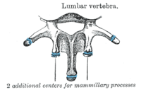
Three portions or tubercles can be noticed in a transverse process of a lower lumbar vertebrae: the lateral or costiform process, the mammillary process, and the accessory process.[2] The costiform is lateral, the mammillary is superior (cranial), and the accessory is inferior (caudal). The mammillary is connected in the lumbar region with the back part of the superior articular process. The accessory process is situated at the back part of the base of the transverse process. The tallest and thickest costiform process is usually that of L5.[2]
First and fifth lumbar vertebrae

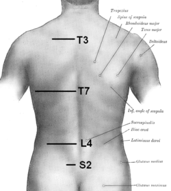
The first lumbar vertebra is level with the anterior end of the ninth rib. This level is also called the important transpyloric plane, since the pylorus of the stomach is at this level. Other important structures are also located at this level, they include; fundus of the gall bladder, celiac trunk, superior mesenteric artery, termination of spinal cord, beginning of fillum terminalis, renal vessels, middle suprarenal arteries, and hila of kidneys.
The fifth lumbar vertebra is characterized by its body being much deeper in front than behind, which accords with the prominence of the sacrovertebral articulation; by the smaller size of its spinous process; by the wide interval between the inferior articular processes, and by the thickness of its transverse processes, which spring from the body as well as from the pedicles.[1] The fifth lumbar vertebra is by far the most common site of spondylolysis and spondylolisthesis.[3]
Most individuals have five lumbar vertebrae, while some have four or six. Lumbar disorders that normally affect L5 will affect L4 or L6 in these latter individuals.
Segmental movements
The range of segmental movements in a single segment is difficult to measure clinically, not only because of variations between individuals, but also because it is age and gender dependent. Furthermore, flexion and extension in the lumbal spine is the product of a combination of rotation and translation in the sagittal plane between each vertebra.[4]
Ranges of segmental movements in the lumbal spine (White and Panjabi, 1990) are (in degrees): [5]
| L1-L2 | L2-L3 | L3-L4 | L4-L5 | L5-S1 | |
|---|---|---|---|---|---|
| Flexion/ Extension | 12° | 14° | 15° | 16° | 17° |
| Lateral flexion | 6° | 6° | 8° | 6° | 3° |
| Axial rotation | 2° | 2° | 2° | 2° | 1° |
Variation

Lumbarization is an anomaly in the spine. It is defined by the nonfusion of the first and second segments of the sacrum. The lumbar spine subsequently appears to have six vertebrae or segments, not five. This sixth lumbar vertebra is known as a transitional vertebra. Conversely the sacrum appears to have only four segments instead of its designated five segments. Lumbosacral transitional vertebrae consist of the process of the last lumbar vertebra fusing with the first sacral segment. [6] While only around 10 percent of adults have a spinal abnormality due to genetics, a sixth lumbar vertebra is one of the more common abnormalities. [7]
Sacralization of the fifth lumbar vertebra (or sacralization) is a congenital anomaly, in which the transverse process of the last lumbar vertebra (L5) fuses to the sacrum on one side or both, or to ilium, or both. These anomalies are observed at about 3.5 percent of people, and it is usually bilateral but can be unilateral or incomplete (ipsilateral or contralateral rudimentary facets) as well. Although sacralization may be a cause of low back pain, it is asymptomatic in many cases (especially bilateral type). Low back pain in these cases most likely occurs due to chronic faulty biomechanics. In sacralization, the L5-S1 intervertebral disc may be thin and narrow. This abnormality is found by X-ray.
Sacralization of L6 means L6 attaches to S1 via a rudimentary joint. This L6-S1 joint creates additional motion, increasing the potential for motion-related stress and lower back pain/conditions. This condition can usually be treated without surgery, injecting steroid medication at the pseudoarticulation instead. Additionally, if L6 fuses to another vertebra this is increasingly likely to cause lower back pain. [8] The presence of a sixth vertebra in the space where five vertebrae normally reside also decreases the flexibility of the spine and increases the likelihood of injury. [9]
Other animals
African apes have three and four lumbar vertebrae, (bonobos have longer spines with an additional vertebra) and humans normally five. This difference, and because the lumbar spines of the extinct Nacholapithecus (a Miocene hominoid with six lumbar vertebrae and no tail) are similar to those of early Australopithecus and early Homo, it is assumed that the Chimpanzee-human last common ancestor also had a long vertebral column with a long lumbar region, and that the reduction in the number of lumbar vertebrae evolved independently in each ape clade. [10] The limited number of lumbar vertebrae in chimpanzees and gorillas result in an inability to lordose (curve) their lumbar spines, in contrast to the spines of Old World monkeys and Nacholapithecus and Proconsul, which suggests that the last common ancestor was not "short-backed" as previously believed. [11]
Additional images
|


See also
References
This article incorporates text in the public domain from the 20th edition of Gray's Anatomy (1918)
- 1 2 3 4 5 6 7 8 Gray's Anatomy (1918), see infobox
- 1 2 Postacchini, Franco (1999) Lumbar Disc Herniation p.19
- ↑ Eizenberg, N. et al. (2008). General Anatomy: Principles and Applications, p. 17.
- ↑ Hansen; et al. (2006). "Anatomy and Biomechanics of the Back Muscles in the Lumbar Spine With Reference to Biomechanical Modeling". Medscape. Archived from the original on November 14, 2010.
- ↑ Hansen; et al. (2006). "Ranges of Segmental Motion for the Lumbar Spine". Medscape. Archived from the original on July 5, 2015.
- ↑ "International Journal of Spine Surgery". Retrieved November 27, 2016.
- ↑ "Dorland's Medical Dictionary". Retrieved November 12, 2008.
- ↑ "Spinal Cord, Inc". Retrieved November 27, 2016.
- ↑ "Laser Spine Institute". Retrieved November 27, 2016.
- ↑ McCollum, MA; Rosenman, BA; Suwa, G; Meindl, RS; Lovejoy, CO (March 15, 2010). "The vertebral formula of the last common ancestor of African apes and humans". J Exp Zool B Mol Dev Evol. 314 (2): 123–34. doi:10.1002/jez.b.21316. PMID 19688850. (Abstract)
- ↑ Lovejoy, C. Owen; McCollum, Melanie A. (October 27, 2010). "Spinopelvic pathways to bipedality: why no hominids ever relied on a bent-hip-bent-knee gait". Philos Trans R Soc Lond B Biol Sci. 365 (1556): 3289–99. doi:10.1098/rstb.2010.0112. PMC 2981964
 . PMID 20855303. (Introduction)
 - ↑ Anatomy Compendium (Godfried Roomans and Anca Dragomir)
External links
| Wikimedia Commons has media related to Lumbar vertebrae. |
- "Lower back pain: Conditions and treatment". SpineUniverse. Retrieved September 2012. Check date values in:
|access-date=(help) - "Virtual Spine — Online Learning Resource". Toronto Western Hospital Departement of Anesthesia. Retrieved September 2012. Check date values in:
|access-date=(help)