Pneumocephalus
| Pneumocephalus | |
|---|---|
 | |
| Pneumocephalus and comminuted fracture of the frontal sinus | |
| Classification and external resources | |
| ICD-10 | G93.2 |
| ICD-9-CM | xxx |
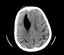
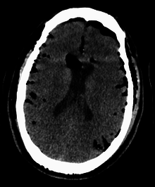
Pneumocephalus is the presence of air or gas within the cranial cavity. It is usually associated with disruption of the skull: after head and facial trauma, tumors of the skull base, after neurosurgery or otorhinolaryngology, and rarely, spontaneously. Pneumocephalus can occur in scuba diving, but is very rare in this context.
If there is a valve mechanism which allows air to enter the skull but prevents it from escaping, a tension pneumocephalus can occur (similar to what can happen in a tension pneumothorax).
CT scans of patients with a tension pneumocephalus typically show air that compresses the frontal lobes of the brain, which results in a tented appearance of the brain in the skull known as the Mount Fuji sign.[1][2][3] The name is derived from the resemblance of the brain to Mount Fuji in Japan, a volcano known for its symmetrical cone. In typical cases, there is a symmetrical depression near the midline (such as the crater of a volcano), due to intact bridging veins.[3] Its occurrence seems to be limited to tension pneumocephalus (not occurring in pneumocephalus without tension).[4] The sign was first described by a team of Japanese neurosurgeons.[5]
Pneumocephalus has also been shown to follow neurosurgical procedures such as deep brain stimulation, where while seemingly innocuous to the patient, may cause brain shift and subsequent stereotactic inaccuracy.[6][7] Efforts are made by neurosurgeons to reduce pneumocephalus volume during surgery, and thus, subsequent brain shift.
Footnotes
- ↑ Michel SJ (August 2004). "The Mount Fuji sign". Radiology. 232 (2): 449–50. doi:10.1148/radiol.2322021556. PMID 15286317.
- ↑ Sadeghian H (September 2000). "Mount Fuji sign in tension pneumocephalus". Archives of Neurology. 57 (9): 1366. doi:10.1001/archneur.57.9.1366. PMID 10987907.
- 1 2 Heckmann JG, Ganslandt O (April 2004). "Images in clinical medicine. The Mount Fuji sign". The New England Journal of Medicine. 350 (18): 1881. doi:10.1056/NEJMicm020479. PMID 15115834.
- ↑ Ishiwata Y, Fujitsu K, Sekino T, et al. (January 1988). "Subdural tension pneumocephalus following surgery for chronic subdural hematoma". Journal of Neurosurgery. 68 (1): 58–61. doi:10.3171/jns.1988.68.1.0058. PMID 3335913.
- ↑ Ishiwata Y, Fujino H, Kubokura T, Tsubone K, Sekino T, Fujitsu K (April 1987). "[Subdural tension pneumocephalus following surgery of chronic subdural hematoma]". No shinkei geka. Neurological surgery (in Japanese). 15 (4): 419–24. PMID 3614535.
- ↑ Elias, W. Jeffrey; Fu, Kai-Ming; Frysinger, Robert C. (2007-11-01). "Cortical and subcortical brain shift during stereotactic procedures". Journal of Neurosurgery. 107 (5): 983–988. doi:10.3171/JNS-07/11/0983. ISSN 0022-3085.
- ↑ Sharim, J; Pezeshkian, P; DeSalles, A; Pouratian, N (2015-10-01). "Effect of Cranial Window Diameter During Deep Brain Stimulation Surgery on Volume of Pneumocephalus". Neuromodulation: Technology at the Neural Interface. 18 (7): 574–579. doi:10.1111/ner.12328. ISSN 1525-1403.