Photoacoustic imaging
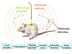
Photoacoustic imaging (optoacoustic imaging) is a biomedical imaging modality based on the photoacoustic effect. In photoacoustic imaging, non-ionizing laser pulses are delivered into biological tissues (when radio frequency pulses are used, the technology is referred to as thermoacoustic imaging). Some of the delivered energy will be absorbed and converted into heat, leading to transient thermoelastic expansion and thus wideband (i.e. MHz) ultrasonic emission. The generated ultrasonic waves are detected by ultrasonic transducers and then analyzed to produce images. It is known that optical absorption is closely associated with physiological properties, such as hemoglobin concentration and oxygen saturation.[1] As a result, the magnitude of the ultrasonic emission (i.e. photoacoustic signal), which is proportional to the local energy deposition, reveals physiologically specific optical absorption contrast. 2D or 3D images of the targeted areas can then be formed.[2] Fig. 1 is a schematic illustration showing the basic principles of photoacoustic imaging.
Biomedical imaging
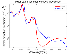
The optical absorption in biological tissues can be due to endogenous molecules such as hemoglobin or melanin, or exogenously delivered contrast agents. As an example, Fig. 2 shows the optical absorption spectra of oxygenated hemoglobin (HbO2) and deoxygenated hemoglobin (Hb) in the visible and near infrared region.[3] Since blood usually has orders of magnitude higher absorption than surrounding tissues, there is sufficient endogenous contrast for photoacoustic imaging to visualize blood vessels. Recent studies have shown that photoacoustic imaging can be used in vivo for tumor angiogenesis monitoring, blood oxygenation mapping, functional brain imaging, skin melanoma detection, methemoglobin measuring, etc.[2]
| Δf | Primary contrast | Δz | δz | δx | Speed | |
|---|---|---|---|---|---|---|
| Hz | mm | µm | µm | Mvx/s | ||
| Photoacoustic microscopy | 50 M | Optical absorption | 3 | 15 | 45 | 0.5 |
| Photoacoustic tomography | 5 M | Optical absorption | 50 | 700 | 700 | 0.5 |
| Confocal microscopy | Fluorescence, scattering | 0.2 | 3-20 | 0.3-3 | 10-100 | |
| Two-photon microscopy | Fluorescence | 0.5-1.0 | 1-10 | 0.3-3 | 10-100 | |
| Optical coherence tomography | 300 T | Optical scattering | 1-2 | 0.5-10 | 1-10 | 20-4.000 |
| Scanning Laser Acoustic Microscopy | 300 M | Ultrasonic scattering | 1-2 | 20 | 20 | 10 |
| Acoustic microscopy | 50 M | Ultrasonic scattering | 20 | 20-100 | 80-160 | 0.1 |
| Ultrasonography | 5 M | Ultrasonic scattering | 60 | 300 | 300 | 1 |
| Table 1. Comparison of contrast mechanisms, penetration depth (Δz), axial resolution (δz), lateral resolution (δx=δy) and imaging speed of confocal microscopy, two-photon microscopy, optical coherence tomography (300 THz), ultrasound microscopy (50 MHz), ultrasound imaging (5 MHz), photoacoustic microscopy (50 MHz), and photoacoustic tomography (3.5 MHz). Speeds in megavoxel per second of non-parallel techniques. | ||||||
Two types of photoacoustic imaging systems, photoacoustic/thermoacoustic computed tomography (also known as photoacoustic/thermoacoustic tomography, i.e., PAT/TAT) and photoacoustic microscopy (PAM), have been developed. A typical PAT system uses an unfocused ultrasound detector to acquire the photoacoustic signals, and the image is reconstructed by inversely solving the photoacoustic equations. A PAM system, on the other hand, uses a spherically focused ultrasound detector with 2D point-by-point scanning, and requires no reconstruction algorithm.
Photoacoustic computed tomography
General equation
Given the heating function , the generation and propagation of photoacoustic wave pressure in an acoustically homogeneous inviscid medium is governed by


where is the speed of sound in medium, is the thermal expansion coefficient, and is the specific heat capacity at constant pressure. Eq. (1) holds under thermal confinement to ensure that heat conduction is negligible during the laser pulse excitation. The thermal confinement occurs when the laser pulsewidth is much shorter than the thermal relaxation time.[4]


The forward solution of Eq. (1) is given by

In stress confinement, which occurs when the laser pulsewidth is much shorter than the stress relaxation time,[4] Eq. (2) can be further derived as
![p({\vec {r}},t)={\frac {1}{4\pi v_{s}^{2}}}{\frac {\partial }{\partial t}}\left[{\frac {1}{v_{s}t}}\int d{\vec {r'}}p_{0}({\vec {r'}})\delta \left(t-{\frac {|{\vec {r}}-{\vec {r'}}|}{v_{s}}}\right)\right]\qquad \,(3),](../I/m/c977afe661f04a20c272d0ec7ca5a61fe45b764e.svg)
where is the initial photoacoustic pressure.

Universal reconstruction algorithm
In a PAT system, the acoustic pressure is detected by scanning an ultrasonic transducer over a surface that encloses the photoacoustic source. To reconstruct the internal source distribution, we need to solve the inverse problem of equation (3) (i.e. to obtain ). A representative method applied for PAT reconstruction is known as the universal backprojection algorithm.[5] This method is suitable for three imaging geometries: planar, spherical, and cylindrical surfaces.
The universal back projection formula is
![\left.p_{0}({\vec {r}})=\int _{{\Omega _{0}}}{\frac {d\Omega _{0}}{\Omega _{0}}}\left[2p({\vec {r_{0}}},v_{s}t)-2v_{s}t{\frac {\partial p({\vec {r_{0}}},v_{s}t)}{\partial (v_{s}t)}}\right]\right|_{{t=|{\vec {r}}-{\vec {r_{0}}}|/v_{s}}},\qquad \quad (4),](../I/m/c656e5723142733bb4ed3b58af7f1e439c4e7b4f.svg)
where is the solid angle subtended by the entire surface with respect to the reconstruction point inside , and


Simple system
A simple PAT/TAT/OAT system is shown in the left part of Fig. 3. The laser beam is expanded and diffused to cover the whole region of interest. Photoacoustic waves are generated proportional to the distribution of optical absorption in the target, and are detected by a single scanned ultrasonic transducer. A TAT/OAT system is the same as PAT except that it uses a microwave excitation source instead of a laser. Although single-element transducers have been employed in these two systems, the detection scheme can be extended to use ultrasound arrays as well.
Biomedical applications
Intrinsic optical or microwave absorption contrast and diffraction-limited high spatial resolution of ultrasound make PAT and TAT promising imaging modalities for wide biomedical applications:
Brain lesion detection
Soft tissues with different optical absorption properties in the brain can be clearly identified by PAT.[6]
Hemodynamics monitoring
Since HbO2 and Hb are the dominant absorbing compounds in biological tissues in the visible spectral range, multiple wavelength photoacoustic measurements can be used to reveal the relative concentration of these two chromophores.[6][7] Thus, the relative total concentration of hemoglobin (HbT) and the hemoglobin oxygen saturation (sO2) can be derived. Therefore, cerebral hemodynamic changes associated with brain function can be successfully detected with PAT.
Breast cancer diagnosis
By utilizing low scattered microwave for excitation, TAT is capable of penetrating thick (several cm) biological tissues with less than mm spatial resolution.[8] Since cancerous tissue and normal tissue have very different responses to radio frequency radiation, TAT has great potential in early breast cancer diagnosis.
Photoacoustic microscopy
The imaging depth of photoacoustic microscopy is mainly limited by the ultrasonic attenuation. The spatial (i.e. axial and lateral) resolutions depend on the ultrasonic transducer used. An ultrasonic transducer with high central frequency and broader bandwidth are chosen to obtain high axial resolution. The lateral resolution is determined by the focal diameter of the transducer. For instance, a 50 MHz ultrasonic transducer provides 15 micrometre axial and 45 micrometre lateral resolution with ~3 mm imaging depth.
Photoacoustic microscopy has multiple important applications in functional imaging: it can detect changes in oxygenated/deoxygenated hemoglobin in small vessels.
See also
References
- ↑ A. Grinvald; et al. (1986). "Functional architecture of cortex revealed by optical imaging of intrinsic signals". Nature. 324 (6095): 361–364. doi:10.1038/324361a0. PMID 3785405.
- 1 2 M. Xu; L.H. Wang (2006). "Photoacoustic imaging in biomedicine". Review of Scientific Instruments. 77 (4): 041101. doi:10.1063/1.2195024.
- ↑ Optical Properties Spectra
- 1 2 L.H. Wang; H.I. Wu (2007). Biomedical Optics. Wiley. ISBN 978-0-471-74304-0.
- ↑ M. Xu; et al. (2005). "Universal back-projection algorithm for photoacoustic-computed tomography". Physical Review E. 71 (1): 016706. doi:10.1103/PhysRevE.71.016706.
- 1 2 X. Wang; et al. (2003). "Non-invasive laser-induced photoacoustic tomography for structural and functional imaging of the brain in vivo". Nature Biotechnology. 21 (7): 803–806. doi:10.1038/nbt839. PMID 12808463.
- ↑ X. Wang; et al. (2006). "Non-invasive imaging of hemoglobin concentration and oxygenation in the rat brain using high-resolution photoacoustic tomography". Journal of Biomedical Optics. 11 (2): 024015. doi:10.1117/1.2192804. PMID 16674205.
- ↑ G. Ku; et al. (2005). "Thermoacoustic and photoacoustic tomography of thick biological tissues toward breast imaging". Technology in Cancer Research and Treatment. 4 (5): 559–566. PMID 16173826.