Pikachurin
| EGFLAM | |||||||||||||||||
|---|---|---|---|---|---|---|---|---|---|---|---|---|---|---|---|---|---|
| Identifiers | |||||||||||||||||
| Aliases | EGFLAM, AGRINL, AGRNL, PIKA, EGF like, fibronectin type III and laminin G domains | ||||||||||||||||
| External IDs | MGI: 2146149 HomoloGene: 65044 GeneCards: EGFLAM | ||||||||||||||||
| |||||||||||||||||
| Orthologs | |||||||||||||||||
| Species | Human | Mouse | |||||||||||||||
| Entrez | |||||||||||||||||
| Ensembl | |||||||||||||||||
| UniProt | |||||||||||||||||
| RefSeq (mRNA) | |||||||||||||||||
| RefSeq (protein) | |||||||||||||||||
| Location (UCSC) | Chr 5: 38.26 – 38.47 Mb | Chr 15: 7.21 – 7.4 Mb | |||||||||||||||
| PubMed search | [1] | [2] | |||||||||||||||
| Wikidata | |||||||||||||||||
| View/Edit Human | View/Edit Mouse |
Pikachurin, also known as AGRINL (AGRINL) and EGF-like, fibronectin type-III and laminin G-like domain-containing protein (EGFLAM), is a protein that in humans is encoded by the EGFLAM gene.[3][4][5]
Pikachurin is a dystroglycan-interacting protein which has an essential role in the precise interactions between the photoreceptor ribbon synapse and the bipolar dendrites.[4] The binding with dystroglycan (DG) depends on several factors (glycosylation of DG, presence of divalent cations, presence of other proteins).
A non-correct binding between pikachurin and DG is associated with muscular dystrophies that often involve eye abnormalities.[6]
Discovery and nomenclature
Pikachurin is an extracellular matrix-like retinal protein first described in 2008 in Japan by Shigeru Sato et al., and named after Pikachu, a character of the Pokémon franchise.[4] The name of this "nimble" protein was inspired due to Pikachu's "lightning-fast moves and shocking electric effects".[7]
Pikachurin was initially identified in a microarray analysis of gene expression profiles of the retinas of wild-type and Otx2 knockout mice. A RT-PCR analysis was used to confirm that Otx2 regulates the expression of pikachurin, it was known because there was an absence of expression of pikachurin in the Otx2 mice retina, so it indicates that Otx2 regulates pikachurin. The localization of pikachurin to synaptic cleft in the photoreceptor ribbon synapse was determined using fluorescent antibodies. Tissue targeting of gene disruption of pikachurin was used to determine that this protein is necessary for proper synaptic signal transmission and visual function. α-dystroglycan was shown to interact with pikachurin through immunoprecipitation.[4]
Pikachurin-dystroglycan interaction
Dystroglycan ligand with other proteins is essential. Glycosylation of dystroglycan is necessary for its ligand binding activity. Mutations in glycosyltransferase enzymes cause abnormal glycosylation of dystroglycan. This hypoglycosylation is associated with less binding with other proteins and causes some congenital muscular dystrophy. Pikachurin is the most recently identified dystroglycan ligand protein and is localized in the synaptic cleft in the photoreceptor ribbon synapse. The binding between dystroglycan and pikachurin requires divalent cations. Ca2+ produces strongest binding; Mn2+ produces only faint bindings and no binding with Mg2+ alone. Dystroglycan has different domains that allow multiple Ca2+ sites to form a stable pikachurin-dystroglycan connection. This shows that pikachurin can form oligomeric structures; and suggests the possibility of clustering effects can be important in modulating pikachurin-dystroglycan interactions. Another thing to be considered is that the presence of NaCl (0.5M) strongly inhibits interaction between DG and other ligand proteins but has a modest inhibitory effect with pikachurin-DG ligand. This shows that there are differences between the binding of pikachurin-DG binding and DG binding with other proteins. Pikachurin seems to have more domains to bind with DG than other proteins. For example, experiments in ligand competition shows that presence of pikachurin inhibits laminin-111 binding with DG, but high concentrations of laminin-111 do not inhibit pikachurin binding to DG.[6]
Function

The protein is colocalized with both dystrophin and dystroglycan at the ribbon synapses.
Pikachurin, along with laminin, perlecan, agrin, neurexin, binds to α-dystroglycan in the extracellular space. As such, pikachurin, as well as the other previously-mentioned proteins, is necessary for the proper functioning of dystroglycan. Pikachurin is necessary for the apposition of presynaptic and postsynaptic termini in the ribbon synapse; deletion of pikachurin causes an abnormal electroretinogram, similarly to the deletion of nestin.[8]
Ribbon synapse relation
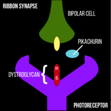
Synapse formation is crucial for the mammalian CNS (central nervous system) to function correctly. Retinal photoreceptors finish at the axon terminal which forms a specialized structure, the ribbon synapse, which specifically connects photoreceptor synaptic terminals with bipolar and horizontal cell terminals in the outer plexiform layer (OPL) of the retina.[4] It is clear that Pikachurin, an extracellular matrix–like retinal protein, is localized to the synaptic cleft in the photoreceptor ribbon synapse.[9] It is demonstrated that with a lack of Pikachurin, there is an improper apposition of the bipolar cell dendritic tips to the photoreceptor ribbon synapses, resulting in alterations in synaptic signal transmission and visual function. The function of Pikachurin remains unknown, but it is a fact that pikachurin is critically involved in the normal photoreceptor ribbon synapse formation and also in physiological functions of visual perception.[10]
Associated pathologies: muscular dystrophies
Congenital muscular dystrophies (CMD) such as muscle-eye-brain disease are caused by defective glycosylation of α-dystroglycan (α-DG) exhibit defective photoreceptor synaptic function. Pikachurin plays an essential role in CMD. Precise interactions between the photoreceptor ribbon synapse and the bipolar dendrites which are realized due to Pikachurin may advance our understanding of the molecular mechanisms underlying the retinal electrophysiological abnormalities observed in muscular dystrophy patients. The muscle-eye-brain dystrophy is caused by mutations in POMGnT1 or LARGE. These two genes mediated a post-translational modification on O-mannose, which is essential for pikachurin binding to dystroglycan, so people who suffer muscle-eye-disease have an hypoglycosylation of pikachurin-α-dystroglycan interactions.[10]
Therapeutic applications
Since pikachurin seems to provide better visual acuity, Sato et al. of the Osaka Bioscience Institute believe that the protein could be used to develop a treatment for retinitis pigmentosa and other eye disorders.[4][11]
See also
- Sonic hedgehog, another protein named after a video game character.
- Zbtb7, an oncogene that was originally named "Pokémon".
- Aerodactylus, a genus of pterosaurs named after a fictional pterosaur in the Pokémon franchise.
References
- ↑ "Human PubMed Reference:".
- ↑ "Mouse PubMed Reference:".
- ↑ "Entrez Gene: EGF-like".
- 1 2 3 4 5 6 Sato S, Omori Y, Katoh K, Kondo M, Kanagawa M, Miyata K, Funabiki K, Koyasu T, Kajimura N, Miyoshi T, Sawai H, Kobayashi K, Tani A, Toda T, Usukura J, Tano Y, Fujikado T, Furukawa T (August 2008). "Pikachurin, a dystroglycan ligand, is essential for photoreceptor ribbon synapse formation". Nat. Neurosci. 11 (8): 923–31. doi:10.1038/nn.2160. PMID 18641643.
- ↑ Gu XH, Lu Y, Ma D, Liu XS, Guo SW (October 2009). "[Model of aberrant DNA methylation patterns and its applications in epithelial ovarian cancer.]". Zhonghua Fu Chan Ke Za Zhi (in Chinese). 44 (10): 754–9. PMID 20078962.
- 1 2 Kanagawa M, Omori Y, Sato S, Kobayashi K, Miyagoe-Suzuki Y, Takeda S, Endo T, Furukawa T, Toda T (October 2010). "Post-translational maturation of dystroglycan is necessary for pikachurin binding and ribbon synaptic localization". J. Biol. Chem. 285 (41): 31208–16. doi:10.1074/jbc.M110.116343. PMC 2951195
 . PMID 20682766.
 - ↑ Levenstein, Steve (2008-07-24). "Lightning-Fast Vision Protein Named After Pikachu". Inventor Spot. Retrieved 2008-07-29.
- ↑ Satz JS, Philp AR, Nguyen H, Kusano H, Lee J, Turk R, Riker MJ, Hernández J, Weiss RM, Anderson MG, Mullins RF, Moore SA, Stone EM, Campbell KP (October 2009). "Visual impairment in the absence of dystroglycan". J. Neurosci. 29 (42): 13136–46. doi:10.1523/JNEUROSCI.0474-09.2009. PMC 2965532. PMID 19846701.
- ↑ Jakob S Satz; Kevin P Campbell (August 2008). "Unraveling the Ribbon Synapse". Nature Neuroscience. 11 (8): 857–859. doi:10.1038/nn0808-857.
- 1 2 Hu H, Li J, Zhang Z, Yu M (February 2011). "Pikachurin interaction with dystroglycan is diminished by defective O-mannosyl glycosylation in congenital muscular dystrophy models and rescued by LARGE overexpression". Neurosci. Lett. 489 (1): 10–5. doi:10.1016/j.neulet.2010.11.056. PMC 3018538. PMID 21129441.
- ↑ "Researchers: 'Pikachurin' protein linked with kinetic vision". Yomiuri Shimbun. 2008-07-22. Archived from the original on 2008-07-27. Retrieved 2008-07-22.
External links
- Lightning-Fast Vision Protein Named After Pikachu - July 24, 2008