Pseudarthrosis
| Pseudarthrosis | |
|---|---|
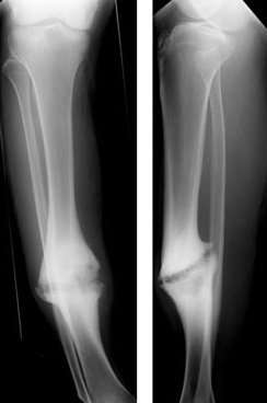 | |
| Hypertrophic nonunion of the tibia | |
| Classification and external resources | |
| Specialty | rheumatology |
| ICD-10 | M84.1, M96.0 |
| MeSH | C21.866.404.468.627 |
Pseudarthrosis (commonly referred to as a nonunion or false joint) is a bone fracture that has no chance of mending without intervention. In pseudarthrosis the body perceives bone fragments as separate bones and does not attempt to unite them. Often this is the result of inadequate healing after the fracture, though it can also be the result of a developmental failure. In the U.S., FDA guidelines allow a period of 9 months for fracture union, after which intervention is considered to facilitate union.
Etymology
The Greek stem "pseudo-" means false and "arthrosis" means joint. Pseudarthrosis, then, is a false joint. In the case of a "non-union", a fracture that does not heal, this is often evidenced by the obliteration of the medullary cavity of a long bone at the site of the non union. This non union is not a true joint, and has no ligamentous support, but often has motion, and remodels into a rounded end that looks like a true joint.
Causes of Nonunion
- Causes related to patient:
- Age: Common in old age
- Nutritional status : poor
- Habits : Nicotine and alcohol consumption
- Metabolic disturbance : Hyperparathyroidism
- Causes related to fracture:
- Distraction at fracture site
- Soft tissue interposition
- Bone loss at the fracture
- Infection
- Loss of blood supply
- Damage of surrounding muscles
- Causes related to treatment
- Inadequate reduction
- Insufficient immobilization
- Improperly applied fixation devices.
- can be found in those with NF1
Types of Nonunion
Judet and Judet, Muller, Weber and Cech, and others classified nonunions into two types according to the viability of the ends of the fragments: Hypervascular nonunions and avascular nonunions.
Hypervascular nonunions are subdivided as:
- "Elephant foot" nonunions: These are hypertrophic, rich in callus and are a result of inadequate immobilisation, insecure fixation or premature weight bearing.
- "Horse hoof" nonunions: Mildly hypertrophic, poor in callus and is due to unstable fixation.
- Oligotrophic nonunions: They are not hypertrophic but vascular, no callus seen and is due to severely displaced fracture or fixation without accurate apposition of fragments.
Avascular nonunions are subdivided as:
- Torsion wedge nonunions have an intermediate fragment with decreased or absent blood supply. This fragment has healed to one main fragment but not to the other.
- Comminuted nonunions have one or more intermediate fragments that are necrotic.
- Defect nonunions has a gap in diaphysis of bone due to a loss of fragment.
- Atrophic nonunions usually are the final result when the intermediate fragments are missing and scar tissue that lacks osteogenic potential is left in their place.
Paley classified tibial nonunions based on clinical and roentgenographic characteristics as Type A (Bone loss of less than 1 cm) and Type B (Bone loss of more than 1 cm). Type A is subclassified as Type A:1 Lax type; Lax nonunion have limited mobility and usually some fixed deformity, Type A:2:1 stiff nonunion without deformity and Type A:2:2 stiff nonunion with a deformity. Type B subclassified as Type B:1 bony defect with no shortening, Type B:2 shortening with no gap and Type B:3 there is both gap and shortening.
Treatment
The point of movement can be treated with electrical stimulations that hopefully will trigger the bone cells to form the hydroxyapatite structure that keeps bones from bending too much. More recently, non-unions are treated by bone grafting, internal fixation, and external fixation, including a technique pioneered by Ilizarov, used to compress the bones at the site of the fracture.[1] Ilizarov originally used bicycle spokes; the modern Taylor Spatial Frame is similar.
Grafting
Donor bone or autologous bone (harvested from the same person undergoing the surgery) is used as a stimulus to bone healing. The presence of the bone is thought to cause stem cells in the circulation and marrow to form cartilage, which then turns to bone, instead of a fibrous scar that forms to heal all other tissues of the body. Bone is the only tissue that can heal without a fibrous scar.
Fixation
Metal plates, pins, screws, and rods, that are screwed or driven into a bone, are used to stabilize the broken bone fragments.
References
External links
| Wikimedia Commons has media related to Pseudarthrosis. |