Quantitative phase-contrast microscopy
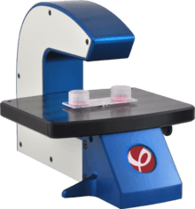 A quantitative phase contrast microscope imaging cultured cells in a slide based cell culture vessel. | |
| Acronym | QPCM |
|---|---|
| Other names | Phase microscope, Quantitative phase microscopy |
| Uses | Microscopic observation and quantification of unstained biological material |
| Related items | Phase contrast microscopy, Differential interference contrast microscopy, Hoffman modulation contrast microscopy, Ptychography |
Quantitative phase contrast microscopy is the collective name for a group of microscopy methods that quantify the phase shift that occurs when light waves pass through a more optically dense object.

Translucent objects, like a living human cell, absorb and scatter small amounts of light. This makes translucent objects difficult to observe in ordinary light microscopes. Such objects do, however, induce a phase shift that can be observed using a phase contrast microscope. Conventional phase contrast microscopy and related methods, such as differential interference contrast microscopy, visualize phase shifts by transforming phase shift gradients into intensity variations. These intensity variations are mixed with other intensity variations, making it difficult to extract quantitative information.[2][3]
Quantitative phase contrast methods are distinguished from other phase contrast methods in that they create a second so-called phase shift image or phase image, independent of the intensity (bright field) image. Phase unwrapping methods are generally applied to the phase shift image to give absolute phase shift values in each pixel, as exemplified by Figure 1.
The principal methods for measuring and visualizing phase shifts include ptychography,[4] Fourier ptychography,[5] and various types of holographic microscopy methods such as digital holographic microscopy, holographic interference microscopy and digital in-line holographic microscopy.[6] Common to these methods is that an interference pattern (hologram) is recorded by a digital image sensor. From the recorded interference pattern, the intensity and the phase shift image is numerically created by a computer algorithm.[7]
Conventional phase contrast microscopy is primarily used to observed unstained living cells.[8] Contrary to conventional phase contrast images, phase shift images of living cells are suitable to be processed by image analysis software. This has led to the development of non-invasive live cell imaging and automated cell culture analysis systems based on quantitative phase contrast microscopy.[6][9][10][11] [12]
See also
- Cytometry
- Digital holographic microscopy
- Holographic interference microscopy
- Live cell imaging
- Phase-contrast microscopy
- Ptychography
- Time stretch quantitative phase imaging
References
- ↑ Manuel Kemmler; Markus Fratz; Dominik Giel; Norbert Saum; Albrecht Brandenburg; Christian Hoffmann (2007). "Noninvasive time-dependent cytometry monitoring by digital holography". J. of Biomedical Optics. 12 (6): 064002. Bibcode:2007JBO....12f4002K. doi:10.1117/1.2804926. PMID 18163818.
- ↑ Etienne Cuche; Frédéric Bevilacqua; Christian Depeursinge (1999). "Digital holography for quantitative phase-contrast imaging". Optics Letters. 24 (5): 291–293. Bibcode:1999OptL...24..291C. doi:10.1364/OL.24.000291.
- ↑ Pierre Marquet; Benjamin Rappaz; Pierre J. Magistretti; Etienne Cuche; Yves Emery; Tristan Colomb; Christian Depeursinge (2005). "Digital holographic microscopy: a noninvasive contrast imaging technique allowing quantitative visualization of living cells with subwavelength axial accuracy". Optics Letters. 30 (5): 468–470. Bibcode:2005OptL...30..468M. doi:10.1364/OL.30.000468.
- ↑ Marrison, Joanne; Räty, Marriott, O'Toole (6 August 2013). "Ptychography – a label free, high-contrast imaging technique for live cells using quantitative phase information". Scientific Reports. 3 (2369). Bibcode:2013NatSR...3E2369M. doi:10.1038/srep02369. Cite uses deprecated parameter
|coauthors=(help) - ↑ Zheng, Guoan; R. Horstmeyer, C. Yang (29 July 2013). "Wide-field, high-resolution Fourier ptychographic microscopy". Nature Photonics. 7. doi:10.1038/nphoton.2013.187. Cite uses deprecated parameter
|coauthors=(help) - 1 2 "4Deep inwater imaging".
- ↑ Myung K. Kim (2010). "Principles and techniques of digital holographic microscopy". SPIE Reviews. 1: 018005. Bibcode:2010SPIER...1a8005K. doi:10.1117/6.0000006.
- ↑ "The phase-contrast microscope". Nobel Media AB.
- ↑ Popescu, G.; Ikeda, T.; Dasari, R. R.; Feld, M. S. (2006). "Diffraction phase microscopy for quantifying cell structure and dynamics". Optics Letters. 31 (6): 775–777. Bibcode:2006OptL...31..775P. doi:10.1364/OL.31.000775. PMID 16544620.
- ↑ "Quantitative phase contrast imaging microscopy - label-free live cell imaging and analysis". Phase Holographic Imaging AB.
- ↑ "Ovizio Imaging Systems".
- ↑ Chen, Claire Lifan; Mahjoubfar, Ata; Tai, Li-Chia; Blaby, Ian K.; Huang, Allen; Niazi, Kayvan Reza; Jalali, Bahram (2016). "Deep Learning in Label-free Cell Classification". Scientific Reports. Full text download available. 6: 21471. Bibcode:2016NatSR...621471C. doi:10.1038/srep21471. PMC 4791545
 . PMID 26975219.published under CC BY 4.0 licensing
External links
- Phase shift time-lapse microscopy video of a triploid cell division
- Cell identification with computational 3D holographic microscopy
Optical microscopy | ||
|---|---|---|
| Illumination and contrast methods |  | |
| Fluorescence methods | ||
| Sub-diffraction limit techniques | ||