Renal column
| Renal column | |
|---|---|
 Kidney | |

| |
| Details | |
| System | Urinary system |
| Identifiers | |
| Latin | columnae renales |
| TA | A08.1.01.019 |
| FMA | 17633 74029, 17633 |
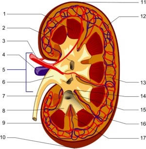
The renal column (or Bertin column, or column of Bertin) is a medullary extension of the renal cortex in between the renal pyramids. It allows the cortex to be better anchored.
Each column consists of lines of blood vessels and urinary tubes and a fibrous material.
A hypertrophied renal column (or renal pseudotumor) may be differentiated from an actual renal tumor with the help of a DMSA scan. The scan will show the area as one with normal activity if it is a pseudotumor or will show decreased uptake if it is a cystic or solid renal mass.
See also
Additional Images
- Renal column
- Renal column
External links
- Anatomy photo:40:06-0106 at the SUNY Downstate Medical Center - "Posterior Abdominal Wall: Internal Structure of a Kidney"
- 01055 at CHORUS
- Histology image: 15901loa – Histology Learning System at Boston University - "Urinary System: neonatal kidney"
- Image at mgh.harvard.edu
This article is issued from Wikipedia - version of the 12/4/2016. The text is available under the Creative Commons Attribution/Share Alike but additional terms may apply for the media files.