Shin splints
| Medial Tibial Stress Syndrome | |
|---|---|
|
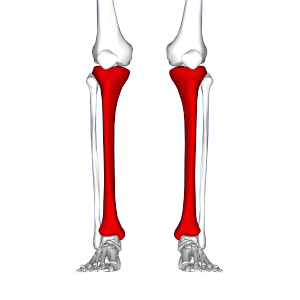
Red area represents tibia. MTSS pain found on inner and lower 2/3rds of tibia. | |
| Classification and external resources | |
| Specialty | Sports medicine |
| Patient UK | Medial Tibial Stress Syndrome |
Shin splints, also known as medial tibial stress syndrome (MTSS), is defined by the American Academy of Orthopaedic Surgeons as "pain along the inner edge of the shinbone (tibia)."[1] Shin splints are usually caused by repeated trauma to the connective muscle tissue surrounding the tibia. They are a common injury affecting athletes who engage in running sports or other forms of physical activity, including running and jumping. They are characterized by general pain in the lower region of the leg between the knee and the ankle. Shin splints injuries are specifically located in the middle to lower thirds of the outside or lateral part of the tibia, which is the larger of two bones comprising the lower leg.
Shin splints are the most prevalent lower leg injury[2] and affect a broad range of individuals. It affects mostly runners and accounts for approximately 13% to 17% of all running-related injuries.[3][4] High school age runners see shin splints injury rates of approximately 13%.[5] Aerobic dancers have also been known to suffer from shin splints, with injury rates as high as 22%.[6] Military personnel undergoing basic training experience shin splints injury rates between 4%-6.4%[7] and 7.9%.[8]
Signs and symptoms
Shin splint pain is described as a recurring dull ache along the inner part of the lower two-thirds of the tibia.[9] In contrast, stress fracture pain is localized to the fracture site.[10]
Biomechanically, over-pronation is the common cause for shin splints and action should be taken to offset the biomechanical irregularity.[11] Pronation occurs when the ankle bone moves downward and towards the middle to create a more stable point of contact with the ground.[12] In other words, the ankle rolls inwards so that more of the arch has contact with the ground. This abnormal movement causes muscles to fatigue more quickly and to be unable to absorb any shock from the foot hitting the ground.[13]
Causes
While the exact cause is unknown, shin splints can be attributed to the overloading of the lower leg due to biomechanical irregularities resulting in an increase in stress exerted on the tibia. A sudden increase in intensity or frequency in activity level fatigues muscles too quickly to properly help absorb shock, forcing the tibia to absorb most of that shock. This stress is associated with the onset of shin splints.[14] Muscle imbalance, including weak core muscles, inflexibility and tightness of lower leg muscles, including the gastrocnemius, soleus, and plantar muscles (commonly the flexor digitorum longus) can increase the possibility of shin splints.[15] The pain associated with shin splints is caused from a disruption of Sharpey's fibres that connect the medial soleus fascia through the periosteum of the tibia where it inserts into the bone.[14] With repetitive stress, the impact forces eccentrically fatigue the soleus and create repeated tibial bending or bowing, contributing to shin splints. The impact is made worse by running uphill, downhill, on uneven terrain, or on hard surfaces. Improper footwear, including worn-out shoes, can also contribute to shin splints.[16][17]
Diagnosis
Shin splints can be diagnosed by a physician after taking a thorough history and performing a complete physical examination. The physical examination focuses on palpable, or gentle pressure, tenderness over a 4–6 inch section on the lower, inside shin area.[18] The pain has been described as a dull ache to an intense pain that increases during exercise, and some individuals experience swelling in the pain area.[19] Clinical history focuses on an individual's previous history with shin splints. People who have previously had shin splints are more likely to have it again.[18]
Vascular and neurological examinations produce normal results in patients with shin splints. Radiographies and three-phase bone scans are recommended to differentiate between shin splints and other causes of chronic leg pain. Bone sctintigraphy and MRI scans can be used to differentiate between stress fractures and shin splints.[18]
It is important to differentiate between different lower leg pain injuries, including shin splints, stress fractures, compartment syndrome, nerve entrapment, and popliteal artery entrapment syndrome. These conditions often have many overlapping symptoms which makes a final diagnosis difficult, and correct diagnosis is needed to determine the most appropriate treatment.
If shin splints are not treated properly, or if exercise is resumed too early or aggressively, shin splints can become permanent.[17]
Treatment
Typical treatments include rest, ice, strengthening and gradually returning to activity.[15] Rest and ice work to allow the tibia to recover from sudden, high levels of stress and reduce inflammation and pain levels. It is important to significantly reduce any pain or swelling before returning to activity. Strengthening exercises should be performed after pain has subsided, on lower leg and hip muscles.[15] Individuals should gradually return to activity, beginning with a short and low intensity level. Over multiple weeks, they can slowly work up to normal activity level. It is important to decrease activity level if any pain returns. Individuals should consider running on other surfaces besides asphalt, such as grass, to decrease the amount of force the lower leg must absorb.[13] Orthoses and insoles help to offset biomechanical irregularities, like pronation, and help to support the arch of the foot.[20] Other conservative interventions include footwear refitting, orthotics, manual therapy, balance training (eg. using a balance board), cortisone injections, and calcium and vitamin D supplementation.[15]
Less common forms of treatment for more severe cases of shin splints include extracorporeal shockwave therapy (ESWT) and surgery.[21] Surgery is only performed in extreme cases where more conservative options have been tried for at least a year.[22] However, surgery does not guarantee 100% recovery.
Epidemiology
Risk factors for developing shin splints include:
- Excessive pronation at subtalar joint[2]
- Excessively tight calf muscles (which can cause excessive pronation)[23]
- Engaging the medial shin muscle in excessive amounts of eccentric muscle activity[2]
- Undertaking high-impact exercises on hard, noncompliant surfaces (ex: running on asphalt or concrete)[2]
- Smoking and low fitness level[24]
While medial tibial stress syndrome is the most common form of shin splints, compartment syndrome and stress fractures are also common forms of shin splints. Females are 1.5 to 3.5 times more likely to progress to stress fractures from shin splints.[2][5][25] This is due in part to females having a higher incidence of diminished bone density and osteoporosis.
References
- ↑ "Shin Splints-OrthoInfo - AAOS".
- 1 2 3 4 5 Yates, B., White, S. (2004). The incidence and risk factors in the development of medial tibial stress syndrome among naval recruits. American Journal of Sports Medicine, 32(3), 772–780.
- ↑ Clement D., Taunton J., Smart G. (1981). "A survey of overuse running injuries". The Physician and Sports Medicine 9, 47–58.
- ↑ Cox J. S., Lenz H. W. (1984). Women midshipmen in sports. American Journal of Sports Medicine, 12(3), 241 – 243.
- 1 2 Bennett J. E.Reinking M. F., Pluemer B., et al. (2001). Factors contributing to the development of medial tibial stress syndrome in high school runners. This could cause pain along the shin. Journal of Orthopedic and Sports Physical Therapies, 31, 504–510.
- ↑ Taunton J. E., McKenzie D. C., Clement D. B. (1988). "The role of biomechanics in the epidemiology of injuries". Sports Medicine, 6, 107–120.
- ↑ Almeida S., Trone D., Leone D., Shaffer R., et al. (1999) "Gender differences in musculoskeletal injury rates: A function of symptom reporting?". Medicine and Science in Sports and Exercise, 31, 1807–1812.
- ↑ Sharma, J., Golby, J., Greeves, J., Spears, I. (2011). "Biomechanical and lifestyle risk factors for medial tibial stress syndrome in army recruits: A prospective study". Gait & Posture, 33,361-365).
- ↑ Carr, K., & Sevetson, E. (2008). Shin splints are also known as shin splinters due to tiny fragments of bone irritating leg muscles. How can you help athletes prevent and treat shin splints? Journal of Family Practice, 57(6), 406–408. Retrieved from EBSCOhost.
- ↑ Edwards Jr., P. H., Wright, M. L., & Hartman, J. F. (2005). A Practical Approach for the Differential Diagnosis of Chronic Leg Pain in the Athlete.American Journal of Sports Medicine, 33(8), 1241–1249.
- ↑ Sharma, J., Golby, J., Greeves, J., Spears, I. (2011). Biomechanical and lifestyle risk factors for medial tibial stress syndrome in army recruits: A prospective study. Gait & Posture, 33,361-365).
- ↑ Sommer, H. & Vallentyne, S. Effect of foot posture on the incidence of medial tibial stress syndrome. Medicine and Science in Sports & Exercise 1995; 800-804
- 1 2 Yates, B., & White, S. The incidence and risk factors in the development of medial tibial stress syndrome among naval recruits. The American Journal of Sports Medicine 2004; 32.3: 772-780
- 1 2 Craig, D. I. (2008). "Medial Tibial Stress Syndrome: Evidence-Based Prevention". Journal of Athletic Training 43(3), 316–318. Retrieved from EBSCOhost.
- 1 2 3 4 Galbraith, R. Michael; Lavallee, Mark E. (2009-10-07). "Medial tibial stress syndrome: conservative treatment options". Current Reviews in Musculoskeletal Medicine. 2 (3): 127–133. doi:10.1007/s12178-009-9055-6. ISSN 1935-973X. PMC 2848339
 . PMID 19809896.
 - ↑ "Running 101: How To Select The Best Pair Of Running Shoes - Competitor.com". 9 September 2014.
- 1 2 "Shin Splints Symptoms, Treatment, Recovery, and Prevention from WebMD".
- 1 2 3 Moen, M., Tol, J., Weir, A., Steunebrink, M. & De Winter, T. Medial tibial stress syndrome: a critical review. Sports Med 2009; 39.7: 524-544
- ↑ Tweed, J., Avil, S., Campbell, J., & Barnes, M. Etiologic factors in the development of medial tibial stress syndrome. Journal of the American Podiatric Medical Association2008; 98.2: 107-112
- ↑ Loudon J. & Dolphino, M. Use of foot orthoses and calf stretching for individuals with medial tibial stress syndrome. Foot Ankle Spec. 2010; 3.1: 15-20
- ↑ Rompe, J., Cacchio, A., Furia, J., & Maffulli, N. Low-energy extracorporeal shock wave therapy as a treatment for medial tibial stress syndrome. The American Journal of Sports Medicine 2010; 38.1:125-132
- ↑ Yates, B., Allen, M., & Barnes, M. Outcome of surgical treatment of medial tibial stress syndrome. The Journal of Bone and Joint Surgery, Incorporated 2003; 85.10: 1974-1980
- ↑ Brukner, P. (2000). Exercise-related lower leg pain: An overview. Medicine and Science in Sports and Exercise, 32(3), pp S1–S3
- ↑ Sharma, J.,Spears, I., Golby, J., Rennie, P., (2009). Medial tibial stress syndrome. 31(1) 45-46
- ↑ Haycock C. E., Gillette J. V. (1976). Susceptibility of women athletes to injury: Myths vs. reality. Journal of the American Medical Association, 236(2), 163–165.
Further reading
- Alonso‐Bartolome, P. (2006). Medial tibial stress syndrome due to methotrexate osteopathy. 65(6): 832–833. Retrieved from Pubmed.
- Haycock C. E., Gillette J. V. (1976). Susceptibility of women athletes to injury: Myths vs. reality. Journal of the American Medical Association, 236(2), 163–165.
- Moen, M., Tol, J., Weir, A., Steunebrink, M. & De Winter, T. Medial tibial stress syndrome: a critical review. Sports Med 2009; 39.7: 524-544
- Raissi, G.R. (2009). The relationship between lower extremity alignment and Medial Tibial Stress Syndrome among non-professional athletes. 1: 11. Retrieved from Pubmed.
- Rompe, J., Cacchio, A., Furia, J., & Maffulli, N. Low-energy extracorporeal shock wave therapy as a treatment for medial tibial stress syndrome. The American Journal of Sports Medicine 2010; 38.1:125-132
- Sommer, H. & Vallentyne, S. Effect of foot posture on the incidence of medial tibial stress syndrome. Medicine and Science in Sports & Exercise 1995; 800-804
- Tweed, J., Avil, S., Campbell, J., & Barnes, M. Etiologic factors in the development of medial tibial stress syndrome. Journal of the American Podiatric Medical Association2008; 98.2: 107-112
- Yates, B., Allen, M. J., & Barnes, M. R. (2003). Outcome of Surgical Treatment of Medial Tibial Stress Syndrome. Journal of Bone & Joint Surgery, American Volume, 85(10), 1974. Retrieved from EBSCOhost.