Separated shoulder
| Separated shoulder | |
|---|---|
| Synonyms | acromioclavicular separation, AC joint separation, AC separation |
.png) | |
| A Separated Shoulder XRay modified to easily show bones. Notice the separation between the end of the collarbone and the scapula. | |
| Classification and external resources | |
| Specialty | orthopedics |
| ICD-10 | S43.1 |
| ICD-9-CM | 831.04, 831.14 |
| eMedicine | orthoped/462 |
A separated shoulder (also known as acromioclavicular separation, AC separation), is a common injury to the acromioclavicular joint. It is a joint dislocation, but it is not called a dislocated shoulder, as that term refers to a glenohumeral joint (shoulder joint) dislocation. The AC joint is located at the distal end of the clavicle, known as the acromial end, and attaches to the acromion of the scapula. Although this is part of the shoulder, a dislocated shoulder and a separated shoulder are completely different. Acromioclavicular separation occurs as a result of a downward force being applied to the superior part of the acromion, either by something striking the top of the acromion or by falling directly on it. The injury is more likely to occur if the shoulder is struck with the hand outstretched. Despite the scapula pulling on the clavicle during impact, the clavicle remains in its general fixed position because of the sternoclavicular joint ligaments.
The trauma to the shoulder affects the ligaments holding the two bones—the scapula and the clavicle—together. This injury does not always involve bone fractures; however if the impact to the shoulder is severe, fractures may occur.
There are four types of soft tissue disruptions that may cause acromioclavicular separation:
- The conoid and trapezoid ligaments may tear at any location
- The lateral clavicle may ride upward after being avulsed from its periosteum
- The acromioclavicular ligaments may be torn
- The conoid-trapezoid ligament origin may avulse from the coracoid
Cause

Separated shoulders often occur in people who participate in sports such as football, soccer, horseback riding, hockey, parkour, combat sports, rowing, rugby, snowboarding, skateboarding, crack the whip, cycling, roller derby and wrestling. The separation is classified into 6 types, with 1 through 3 increasing in severity, and 4 through 6 being the most severe. The most common mechanism of injury is a fall on the tip of the shoulder or also a fall on an outstretched hand. In falls where the force is transmitted indirectly, often only the acromioclavular ligament is affected, and the coracoclavicular ligaments remain unharmed.[1] In ice hockey, the separation is sometimes due to a lateral force, as when one gets forcefully checked into the side of the rink.[2]
Mechanism
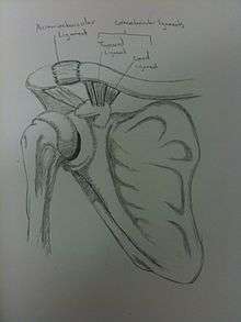
The acromion of the scapula is connected to the clavicle by the superior acromioclavicular ligament. The coracoclavicular ligaments connect the clavicle to the coracoid process. The two ligaments that form the coracoclavicular ligaments are the trapezoid and conoid ligaments. These three ligaments add support to the shoulder joint.
Diagnosis
Diagnosis is based on physical examination and an x-ray. A separated shoulder occurs because of a direct blow to the AC joint or a fall on the elbow that forces the head of the humerus into the AC joint. Furthermore, AC separation can be identified point tenderness, pain at the AC joint with cross-arm adduction, and pain relief with an injection of a local anesthetic. The cross-arm adduction will produce pain specifically at the AC joint and will be done by elevating the arm to a 90° angle, flexing the elbow to a 90° angle, and adducting the arm across the chest. The pain in the shoulder is hard to pinpoint of the innervation of the AC joint and the glenohumeral joint. An injury to the AC joint will result in pain over the AC joint, in the anterolateral neck and in the region in the anterolateral deltoid.
It can be classified into 6 types.
Type I
A Type I AC separation involves direct trauma to the shoulder causing the injury to ligaments that form the joint, but no severe tearing or fracture. It is commonly referred to as a sprain.[3]
Type II
A Type II AC separation involves complete tearing of the acromioclavicular ligament, as well as a partial tear of the coracoclavicular ligaments. This often causes a noticeable bump on the shoulder.[4] This bump is permanent. The clavicle is unstable to direct stress examination. On radiographs, the lateral end of the clavicle may be slightly elevated by pressing on the sternal aspect of the clavicle forcing the acromial end down, and by releasing, it may pop back up eliciting a piano key sign due to the tearing of the AC. Severe pain and loss of movement are common.
Type III

In a Type III AC separation both acromioclavicular and coracoclavicular ligaments are torn without significant disruption of the deltoid or trapezial fascia.[4] A significant bump, resulting in some shoulder deformity, is formed by the lateral end of the clavicle. This bump, caused by the clavicle's dislocation, is permanent. The clavicle can be moved in and out of place on the shoulder. A radiographic examination, will show the results as abnormal. Pain with motion can be severe.
Type IV
This is a type III injury with avulsion of the coracoclavicular ligament from the clavicle, with the distal clavicle displaced posteriorly into or through the trapezius and may tent the posterior skin.[4] A displaced clavicle is easily seen on a radiograph. It is important to evaluate the sternoclavicular joint also, because there can be an anterior dislocation of the sternoclavicular joint and posterior dislocation of the AC joint. This injury is generally acknowledged to require surgery.
Type V
This is a more severe form of a type III injury, with the trapezial and deltoid fascia stripped off of the acromion as well as the clavicle. This is type III but with exaggeration of the vertical displacement of the clavicle from the scapula. Distinguishing between Type III and Type V separations based on radiographs is difficult and often unreliable between surgeons.[5] Type V is manifested by a 2- to 3-fold increase in the coracoclavicular distance.[4] The shoulder manifests as a severe droop, secondary to downward displacement of the scapula and humerus due to loss of the clavicular strut.[4] This injury generally requires surgery.
Type VI
This is type III with inferior dislocation of the distal end of the clavicle below the coracoid. This injury is associated with severe trauma and frequently accompanied by multiple other injuries.[4] The mechanism is thought to be severe hyperabduction and external rotation of the arm, combined with retraction of the scapula. The distal clavicle is found in 2 orientations, either subacromial or subcoracoid. With the subcoracoid dislocation, the clavicle becomes lodged behind the intact conjoined tendon. The posterior superior AC ligaments, which often remains attached to the acromion, get displaced into the AC interval, making anatomic reduction difficult. The tissue needs to be surgically cleared and then reattached after reduction. Most patients with type VI injuries have paresthesia that resolves after relocation of the clavicle [4] It is extremely rare and generally only involved with motor vehicle collisions. This requires surgery.
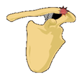 Classification type 1 is the most common type
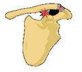 Classification type 2
- Classification type 3
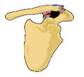 Classification type 4
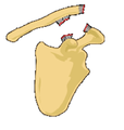 Classification type 5 is rare
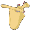 Classification type 6 is rare
Treatment
Treatment of a separated shoulder depends on the severity of the injury. When beginning treatment some of the things one should do first, is control the inflammation, rest the joint, and ice the joint. Take an anti-inflammatory such as Advil or Motrin to help minimize the pain and inflammation. Rest the joint which will also help minimize painful symptoms and allow the healing to begin. When icing, it should be done every four hours for 15 minutes at a time. One can wear a sling until the pain subsides; then some simple exercises can be started.
Non-surgical

Type I and type II shoulder separation are the most common types of separated and rarely need surgery. However, the risk of arthritis with type II separations is greatly increased. If it becomes severe, the Mumford procedure or distal clavicle excision can be performed.
Most non-surgical treatment options include physical therapy to build up the muscles and help stabilize the joint. Literature regarding long-term follow-up after surgical repair of type III injuries is scarce, and those treated nonoperatively generally do quite well. There may also be the potential that surgical repair may be less painful in the long run.
Once the pain has eased, range-of-motion exercises can be started followed by a strength training program. The strength training will include strengthening of the rotator cuff, and shoulder blade muscles. With most cases the pain goes away after three weeks. Although full recovery can take up to six weeks for type II and up to twelve weeks for type III.
Those who do have a separated shoulder will most often return to having full function, although some may have continued pain in the area of the AC joint. With the continued pain there are some things that maybe causing it. It may be due to an abnormal contact between the bone ends when the joint is in motion, the development of arthritis, or an injury to a piece of the cushioning cartilage that is found between the bone ends of this joint.[6]
Surgical
Type IV, V, and VI shoulder separations are very uncommon but always require surgery. There is some debate among orthopedic surgeons, however, about the treatment of type III shoulder separation. Many with type III shoulder separation who do not undergo surgical treatment recover just as well as those who do receive it, and avoid the added risks that surgery may present. Those who opt out of surgery also have faster recovery times and are able to return to work or sports sooner. Some studies suggest early surgical treatment of type III separation may benefit laborers and athletes who perform overhead motions. The potential benefit of surgical treatment for type III remains unproven.[7] Studies have been done that those who did have the surgery or not will need some kind of operation later down the road. The injured joint degenerates a lot faster than normal and over time it can become arthritic and painful.
There have been many surgeries described to fix complete acromioclavicular separations, including recently arthroscopic. There is no consensus on which surgery is best. Several surgeries have been described with pins or hooks. Another surgery performs muscle transfer.
A common surgery is some form of Modified Weaver-Dunn procedure, which involves cutting off the end of the clavicle portion, partially sacrificing the coracoacromial ligament and suturing the displaced acromial end to the lateral aspect of the clavicle for stabilization, then often some form of additional support is introduced to replace the coracoclavicular ligament(s). Variations of this support includes grafting of tendons from the leg. Other surgeries have used a Rockwood screw that is inserted initially and then removed after 12 weeks. Physical therapy is always recommended after surgery, and most patients get flexibility back, although possibly somewhat limited.
After one does have surgery, a sling should be worn to support and protect the shoulder for a few days. For the first couple physical therapy visits, the treatment will focus on controlling the pain and swelling. Type of treatment can include, ice and electrical stimulation, massage, or other hand on treatment to help ease the pain and muscle spasm.
After about four weeks range of motion exercises can be started. Passive exercises are done which the shoulder joint is moved but the muscles stay relaxed. After about six to eight weeks active therapy is started. Such exercises can include isometric strengthening which works the muscles without straining the healing of the joint. After about three months, more active strengthening will be incorporated which focus on improving the strength and control of the rotator cuff muscles and the muscles around the shoulder blade. The exercises that the therapist gives the patient to be done at home should be done to be able to get a better recovery in the long run.[8]
Physical therapy
Some physical therapy exercises that can be performed to help rehab the shoulder are: While standing and using a theraband you can perform Y, T, and W’s, Internal shoulder rotation, External shoulder rotation, Shoulder extensions,and Scapula squeezes While lying on your side you can perform internal rotation and external rotation with a light weight. The light weight can be any type of object such as a 1-5 lb dumbbell weight, or a soup can. Also you can foam roll the pectorals. With the foam roller you can also lie on your back on top of it and do snow angels. [9]
See also
References
- ↑ Gloria M. Beim, MD (July 2000). "Acromioclavicular Joint Injuries". J Athl Train. 35 (3): 261–267. PMC 1323387
 . PMID 16558638.
 - ↑ Stephen Bushee, ATC. "Acromioclavicular Separation in Ice Hockey, Typical injury...different mechanism!". Retrieved 2006-11-01.
- ↑ An examination using radiography (a type of medical imaging) will show up as normal.
- 1 2 3 4 5 6 7 Mazzocca AD, Arciero RA, Bicos J (February 2007). "Evaluation and treatment of acromioclavicular joint injuries". Am J Sports Med. 35 (2): 316–29. doi:10.1177/0363546506298022. PMID 17251175.
- ↑ Kraeutler, MJ; Williams GR Jr; Cohen SB; Ciccotti MG; Tucker BS; Dines JS; Altchek DW; Dodson CC (October 2012). "Inter- and intraobserver reliability of the radiographic diagnosis and treatment of acromioclavicular joint separations". Orthopedics. 35 (10): e1483-1487. doi:10.3928/01477447-20120919-16. PMID 23027484.
- ↑ "Shoulder Separation". OrthoInfo. American Academy of Orthopaedic Surgeons. October 2007.
- ↑ Cluett, Jonathan. "Separated Shoulder". Retrieved 2012-05-05.
- ↑ "Acromioclavicular Joint Separation". ISOST. Retrieved 2010-05-05.
- ↑ http://www.coreperformance.com/knowledge/injury-pain/separated-shoulder.html
External links
| Wikimedia Commons has media related to Separated shoulder. |
- Wheeless Online online orthopedic resource (for orthopedists)
- Arthroscopic Weaver-Dunn
- Rollo J, Raghunath J, Porter K (October 2005). "Injuries of the acromioclavicular joint and current treatment options". Trauma. 7 (4): 217–223. doi:10.1191/1460408605ta349oa.
- Acromioclavicular Injuries: New Management Options Emerge (free with registration)