Sodium MRI
Like more common forms of MRI, sodium MRI relies on the use of a powerful magnetic field to induce alignment of nuclei with magnetic spin.[1][2] 1H nuclei, for example, have a magnetic moment caused by the angular momentum of the charged proton, therefore H2O water molecules can be visualized when in the presence of a strong magnetic field. Although most MRI is done to image 1H nuclei, other nuclei, such as 23Na, can be visualized as well. Because Na+ ions play key roles in biological processes, homeostasis, and metabolism, imaging 23Na nuclei can provide direct insights into the metabolic activity and cellular integrity of tissues.[3] However, 23Na MRI is complicated by the low concentrations of Na nuclei in biological tissues[4] (10-15 mM) as compared to concentration of H2O molecules, and the lower gyromagnetic ratio of the 23Na nucleus as compared to a 1H nucleus[5] (26% of the 1H nucleus gyromagnetic ratio),[6] causing low NMR sensitivity and requiring a stronger magnetic field for higher resolution. The quadrupolar 23Na nucleus has very fast transverse relaxation rates and multiple quantum coherences as compared to the 1H nucleus.[6]
Biological significance
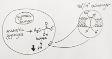
Tissue sodium concentration (TSC) is tightly regulated by healthy cells and are altered by energy status and cellular integrity, making it an effective marker for disease states.[4][6][7] Cells maintain a low intracellular Na+ concentration by actively pumping Na ions out via the Na+/K+ ATPase channel, and any challenge to the cell’s metabolism causing low ATP supply or compromise of the cell’s membrane integrity will drastically increase intracellular Na+ concentrations. After exhaustive exercise, for example, 23Na MRI can detect Na+ levels in tissues rising sharply, and can even visualize a sodium-rich meal in a patient’s stomach. Malignant tumors in particular alter their metabolism drastically, often to account for hypoxic intratumor conditions, leading to an decrease in cytosolic pH. To compensate, Na+ ions from the extracellular space are exchanged for protons in the Na+/H+ antiport,[6] the loss of which often attenuates cancer growth.[8] Therefore, 23Na MRI is a useful clinical tool for detecting a number of disease states, including heart disease[9] and cancer, as well as monitoring therapy. Tissue damage in stroke patients can be evaluated using 23Na MRI, with one study showing that a change of 50% higher TSC than the TSC in healthy brain tissue is consistent with complete infarction,[10] and therefore can be used to determine tissue viability and treatment options for the patient. Tumor malignancy can also be evaluated based on the increases in TSC of rapidly proliferating cells. Malignant tumors have approximately 50-60% increased TSC relative to that of healthy tissues[10] – however, increases in TSC cannot be determined to be due to changes in extracellular volume, intracellular sodium content or neovascularization. Another interesting use of 23Na MRI is in evaluating multiple sclerosis, wherein accumulation of sodium in axons can lead to axon degeneration.[11] Preliminary studies have shown that there is a positive correlation between elevated TSC and disability.
Advantages
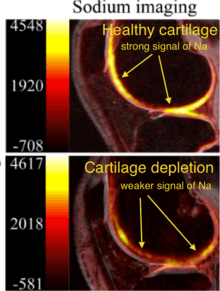
23Na MRI measures cellular metabolic rate as well as disease-related change in tissues and organs.[12] It had improved from 45min length to only 15 mins at 1.5T.[6][10] Unlike other MRI scanning, sodium MRI captures only sodium signals inside bodies. For cartilage degeneration, proteoglycan degrades with negative charge, and positively charged sodium ion bond with proteoglycan.[2] Both proteoglycan and sodium level decrease, so less signals are observed by sodium MRI. 23Na MRI is very sensitive and specific to change in proteoglycan, so it is good to use for monitoring of proteoglycan degeneration in cartilage.[2][13]
References
- ↑ Ouwerkerk, Ronald; Morgan, Russell H. (2007-10-01). "23Na MRI: From Research to Clinical Use". Journal of the American College of Radiology : JACR. 4 (10): 739–741. doi:10.1016/j.jacr.2007.07.001. ISSN 1546-1440. PMC 2084082
 . PMID 17903762.
 - 1 2 3 Borthakur, A; Shapiro, E. M; Beers, J; Kudchodkar, S; Kneeland, J. B; Reddy, R (2000-07-01). "Sensitivity of MRI to proteoglycan depletion in cartilage: comparison of sodium and proton MRI". Osteoarthritis and Cartilage. 8 (4): 288–293. doi:10.1053/joca.1999.0303.
- ↑ Madelin, Guillaume (2012-12-18). "Sodium Magnetic Resonance Imaging: Biomedical Applications". arXiv:1212.4400 [physics.med-ph].
- 1 2 Romanzetti, Sandro; Mirkes, Christian C.; Fiege, Daniel P.; Celik, Avdo; Felder, Jörg; Shah, N. Jon (2014-08-01). "Mapping tissue sodium concentration in the human brain: a comparison of MR sequences at 9.4Tesla". NeuroImage. 96: 44–53. doi:10.1016/j.neuroimage.2014.03.079. ISSN 1095-9572. PMID 24721332.
- ↑ Madelin, Guillaume; Lee, Jae-Seung; Regatte, Ravinder R.; Jerschow, Alexej (2014-05-01). "Sodium MRI: Methods and applications". Progress in Nuclear Magnetic Resonance Spectroscopy. 79: 14–47. doi:10.1016/j.pnmrs.2014.02.001. PMC 4126172. PMID 24815363.
- 1 2 3 4 5 Madelin, Guillaume; Regatte, Ravinder R. (2013-09-01). "Biomedical Applications of Sodium MRI In Vivo". Journal of magnetic resonance imaging : JMRI. 38 (3): 511–529. doi:10.1002/jmri.24168. ISSN 1053-1807. PMC 3759542. PMID 23722972.
- ↑ Ouwerkerk, Ronald (2011-01-01). Modo, Michel; Bulte, Jeff W.M., eds. Sodium MRI. Methods in Molecular Biology. Humana Press. pp. 175–201. ISBN 9781617379918.
- ↑ Alevizopoulos, Konstantinos; Calogeropoulou, Theodora; Lang, Florian; Stournaras, Christos (2014-01-01). "Na+/K+ ATPase inhibitors in cancer". Current Drug Targets. 15 (10): 988–1000. ISSN 1873-5592. PMID 25198786.
- ↑ Bottomley, Paul A. (2016-02-01). "Sodium MRI in human heart: a review". NMR in Biomedicine. 29 (2): 187–196. doi:10.1002/nbm.3265. ISSN 1099-1492. PMID 25683054.
- 1 2 3 Ouwerkerk, Ronald; Bleich, Karen B.; Gillen, Joseph S.; Pomper, Martin G.; Bottomley, Paul A. (2003-05-01). "Tissue Sodium Concentration in Human Brain Tumors as Measured with 23Na MR Imaging". Radiology. 227 (2): 529–537. doi:10.1148/radiol.2272020483. ISSN 0033-8419.
- ↑ Inglese, M.; Madelin, G.; Oesingmann, N.; Babb, J. S.; Wu, W.; Stoeckel, B.; Herbert, J.; Johnson, G. (2010-03-01). "Brain tissue sodium concentration in multiple sclerosis: a sodium imaging study at 3 tesla". Brain. 133 (3): 847–857. doi:10.1093/brain/awp334. ISSN 0006-8950. PMC 2842511. PMID 20110245.
- ↑ Madelin, Guillaume; Kline, Richard; Walvick, Ronn; Regatte, Ravinder R. (2014-04-23). "A method for estimating intracellular sodium concentration and extracellular volume fraction in brain in vivo using sodium magnetic resonance imaging". Scientific Reports. 4. doi:10.1038/srep04763. PMC 4762219. PMID 24755879.
- ↑ Newbould, R.D.; Miller, S.R.; Tielbeek, J.A.W.; Toms, L.D.; Rao, A.W.; Gold, G.E.; Strachan, R.K.; Taylor, P.C.; Matthews, P.M. (2012-01-01). "Reproducibility of sodium MRI measures of articular cartilage of the knee in osteoarthritis". Osteoarthritis and Cartilage. 20 (1): 29–35. doi:10.1016/j.joca.2011.10.007. ISSN 1063-4584. PMC 3270258. PMID 22040861.