Superior hypogastric plexus
| Superior hypogastric plexus | |
|---|---|
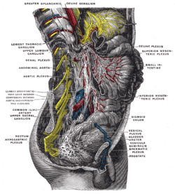
 The right sympathetic chain and its connections with the thoracic, abdominal, and pelvic plexuses. (Hypogastric plexus is labeled on right, fourth from the bottom.) | |
 Lower half of right sympathetic cord. | |
| Details | |
| Identifiers | |
| Latin | plexus hypogastricus superior, nervus presacralis |
| MeSH | A08.800.050.050.400 |
| TA | A14.3.03.046 |
| FMA | 6642 |
The superior hypogastric plexus (in older texts, hypogastric plexus or presacral nerve) is a plexus of nerves situated on the vertebral bodies anterior to the bifurcation of the abdominal aorta.
Structure
From the plexus, sympathetic fibers are carried into the pelvis as two main trunks- the right and left hypogastric nerves- each lying medial to the internal iliac artery and its branches. The right and left hypogastric nerves continues as Inferior hypogastric plexus; these hypogastric nerves send sympathetic fibers to the ovarian and ureteric plexi, which originate within the renal and abdominal aortic sympathetic plexi. The superior hypogastric plexus receives contributions from the two lower lumbar splanchnic nerves (L3-L4), which are branches of the chain ganglia. They also contain parasympathetic fibers which arise from pelvic splanchnic nerve (S2-S4) and ascend from Inferior hypogastric plexus; it is more usual for these parasympathetic fibers to ascend to the left-handed side of the superior hypogastric plexus and cross the branches of the sigmoid and left colic vessel branches, as these parasympathetic branches are distributed along the branches of the inferior mesenteric artery.
Additional images
References
This article incorporates text in the public domain from the 20th edition of Gray's Anatomy (1918)
External links
- Autonomics of the Pelvis - Page 4 of 12 anatomy module at med.umich.edu
- posteriorabdomen at The Anatomy Lesson by Wesley Norman (Georgetown University) (posteriorabdmus&nerves)
- pelvis at The Anatomy Lesson by Wesley Norman (Georgetown University) (pelvicsympathnerves)
- figures/chapter_32/32-6.HTM: Basic Human Anatomy at Dartmouth Medical School
