Thigh
| Thigh | |
|---|---|
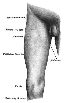 Front and medial aspect of a male right thigh | |
 Cross-section of the thigh showing muscles and bone (latin terminology). | |
| Details | |
| Identifiers | |
| Latin | femur |
| TA | A01.1.00.035 |
| FMA | 24967 |
In humans, the thigh is the area between the pelvis and the knee. Anatomically, it is part of the lower limb.[1]
The single bone in the thigh is called the femur. This bone is very thick and strong (due to the high proportion of cortical bone), and forms a ball and socket joint at the hip, and a modified hinge joint at the knee.
Structure
Bones
The femur is the only bone in the thigh and serves for an attachment site for all muscles in the thigh. The head of the femur articulates with the acetabulum in the pelvic bone forming the hip joint, while the distal part of the femur articulates with the tibia and kneecap forming the knee joint. By most measures the femur is the strongest bone in the body. The femur is also the longest bone in the body.
The femur is categorised as a long bone and comprises a diaphysis, the shaft (or body) and two epiphysis or extremities that articulate with adjacent bones in the hip and knee.[2]
Muscular compartments
In cross-section, the thigh is divided up into three separate compartments, divided by fascia, each containing muscles. These compartments use the femur as an axis, and are separated by tough connective tissue membranes (or septa). Each of these compartments has its own blood and nerve supply, and contains a different group of muscles.
- Medial fascial compartment of thigh, adductor
- Posterior fascial compartment of thigh, extensor, hamstring
- Anterior fascial compartment of thigh, flexor
Anterior compartment muscles of the thigh include pectineus, sartorius, and the four muscles that comprise the quadriceps muscles- rectus femoris, vastus medialis, vastus intermedius and vastus lateralis.
Posterior compartment muscles of the thigh are the hamstring muscles, which include semimembranosus, semitendinosus, and biceps femoris.
Medial compartment muscles are adductor magnus, adductor longus and adductor brevis, and also gracilis.
Because the major muscles of the thigh are the largest muscles of the body, resistance exercises (strength training) of them stimulate blood flow more than any other localized activity. [3]
Blood supply
The arterial supply is by the femoral artery and the obturator artery. The lymphatic drainage closely follows the arterial supply and drains to the lumbar lymphatic trunks on the corresponding side, which in turn drains to the cisterna chyli.
The deep venous system of the thigh consists of the femoral vein, the proximal part of the popliteal vein, and various smaller vessels; these are the site of proximal deep venous thrombosis. The venae perfortantes connect the deep and the superficial system, which consists of the saphenous veins (the site of varicose veins).
Clinical significance
Thigh weakness can result in a positive Gowers' sign on physical examination.
Additional images
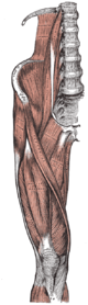 Front of thigh muscles from Gray's Anatomy of the human body from 1918.
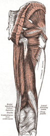 Back thigh muscles of the gluteal and posterior femoral regions from Gray's Anatomy of the human body from 1918.
 Cross-section through the middle of the thigh.
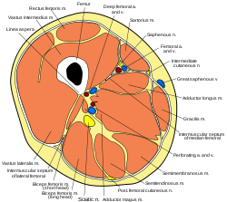 Cross-section through the middle of the thigh.
References
| Look up thigh in Wiktionary, the free dictionary. |
| Wikimedia Commons has media related to Thigh. |
- ↑ "thigh" at Dorland's Medical Dictionary
- ↑ Bojsen-Møller, Finn; Simonsen, Erik B.; Tranum-Jensen, Jørgen (2001). Bevægeapparatets anatomi [Anatomy of the Locomotive Apparatus] (in Danish) (12th ed.). pp. 239–241. ISBN 978-87-628-0307-7.
- ↑ http://www.livestrong.com/article/406641-how-important-are-leg-workouts-for-muscle-gain/