Centriole
| Cell biology | |
|---|---|
| The centrosome | |
|
Components of a typical centrosome:
|
.svg.png)
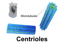
In cell biology a centriole is a cylindrical cell structure[1] composed mainly of a protein called tubulin that is found in most eukaryotic cells. An associated pair of centrioles, surrounded by a shapeless mass of dense material, called the pericentriolar material, or PCM, makes up a compound structure called a centrosome.[1]
Centrioles are present in the cells of most eukaryotes, for example those of animals. However, they are absent from conifers (pinophyta), flowering plants (angiosperms) and most fungi, and are only present in the male gametes of charophytes, bryophytes, seedless vascular plants, cycads, and ginkgo.[2][3]
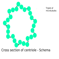
Most centrioles are made up of nine sets of microtubule triplets, arranged in a cylinder. Deviations from this structure include crabs and Drosophila melanogaster embryos, with nine doublets, and Caenorhabditis elegans sperm cells and early embryos, with nine singlets.[4][5]
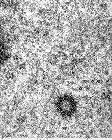
Edouard van Beneden and Theodor Boveri made the first observation and identification of centrioles in 1883 and 1888 respectively,[6][7] while the pattern of centriole duplication was first worked out independently by Etienne de Harven and Joseph G. Gall c. 1950 [8][9] The main function of centrioles is to produce aster and spindle during cell division.
Cell division

Centrioles are involved in the organization of the mitotic spindle and in the completion of cytokinesis.[10] Centrioles were previously thought to be required for the formation of a mitotic spindle in animal cells. However, more recent experiments have demonstrated that cells whose centrioles have been removed via laser ablation can still progress through the G1 stage of interphase before centrioles can be synthesized later in a de novo fashion.[11] Additionally, mutant flies lacking centrioles develop normally, although the adult flies' cells lack flagella and cilia and as a result, they die shortly after birth.[12] The centrioles can self replicate during cell division.
Cellular organization
Centrioles are a very important part of centrosomes, which are involved in organizing microtubules in the cytoplasm.[13][14] The position of the centriole determines the position of the nucleus and plays a crucial role in the spatial arrangement of the cell.
Ciliogenesis
In organisms with flagella and cilia, the position of these organelles is determined by the mother centriole, which becomes the basal body. An inability of cells to use centrioles to make functional cilia and flagella has been linked to a number of genetic and developmental diseases. In particular, the inability of centrioles to properly migrate prior to ciliary assembly has recently been linked to Meckel-Gruber syndrome.[15]
Animal development
Proper orientation of cilia via centriole positioning toward the posterior of embryonic node cells is critical for establishing left–right asymmetry during mammalian development.
Centriole duplication
Before DNA replication, cells contain two centrioles. The older of the two centrioles is termed the mother centriole, the other the daughter. During the cell division cycle, a new centriole grows from each side of the mother centriole. After duplication, the two centriole pairs will remain attached to each other orthogonally until mitosis. At that point the mother and daughter centrioles separate dependently on an enzyme called separase.[16]
The two centrioles in the centrosome are tied to one another. The mother centriole has radiating appendages at the distal end of its long axis and is attached to its daughter at the proximal end. Each daughter cell formed after cell division will inherit one of these pairs. Centrioles start duplicating when DNA replicates.[10]
Origin
The last common ancestor of all eukaryotes was a ciliated cell with centrioles. Some lineages of eukaryotes, such as land plants, do not have centrioles except in their motile male gametes. Centrioles are completely absent from all cells of conifers and flowering plants, which do not have ciliate or flagellate gametes.[17] It is unclear if the last common ancestor had one[18] or two cilia.[19] Important genes required for centriole growth, like centrins, are only found in eukaryotes and not in bacteria or archaeans.[18]
Etymology and pronunciation
The word centriole (/ˈsɛntrioʊl/) uses combining forms of centri- and -ole, yielding "little central part", which describes a centriole's typical location near the center of the cell.
References
- 1 2 Eddé, B; Rossier, J; Le Caer, JP; Desbruyères, E; Gros, F; Denoulet, P (1990). "Posttranslational glutamylation of alpha-tubulin". Science. 247 (4938): 83–5. Bibcode:1990Sci...247...83E. doi:10.1126/science.1967194. PMID 1967194.
- ↑ Quarmby, LM; Parker, JD (2005). "Cilia and the cell cycle?". The Journal of Cell Biology. 169 (5): 707–10. doi:10.1083/jcb.200503053. PMC 2171619
 . PMID 15928206.
 - ↑ Silflow, CD; Lefebvre, PA (2001). "Assembly and motility of eukaryotic cilia and flagella. Lessons from Chlamydomonas reinhardtii.". Plant Physiology. 127: 1500–1507. doi:10.1104/pp.010807. PMC 1540183. PMID 11743094.
- ↑ Delattre, M; Gönczy, P (2004). "The arithmetic of centrosome biogenesis". Journal of Cell Science. 117 (Pt 9): 1619–30. doi:10.1242/jcs.01128. PMID 15075224.
- ↑ Leidel, S; Delattre, M; Cerutti, L; Baumer, K; Gönczy, P (2005). "SAS-6 defines a protein family required for centrosome duplication in C. elegans and in human cells". Nature Cell Biology. 7 (2): 115–25. doi:10.1038/ncb1220. PMID 15665853.
- ↑ Wunderlich, V. (2002). "JMM - Past and Present". Journal of Molecular Medicine. 80 (9): 545–548. doi:10.1007/s00109-002-0374-y. PMID 12226736.
- ↑ Boveri, Theodor (2012). Zellen-Studien II: Die Befruchtung und Teilung des Eies von Ascaris megalocephala. Jena: Gustav Fischer Verlag.
- ↑ Wolfe, Stephen L. (1977). Biology: the foundations (First ed.). Wadsworth Pub. Co.
- ↑ Vorobjev, I. A.; Nadezhdina, E. S. (1987). "The Centrosome and Its Role in the Organization of Microtubules". International Review of Cytology. International Review of Cytology. 106: 227–293. doi:10.1016/S0074-7696(08)61714-3. ISBN 978-0-12-364506-7. PMID 3294718.. See also de Harven's own recollections of this work: de Harven, Etienne (1994). "Early observations of centrioles and mitotic spindle fibers by transmission electron microscopy" (PDF). Biol Cell. 80 (2–3): 107–109. doi:10.1016/0248-4900(94)90028-0. PMID 8087058.
- 1 2 Salisbury, JL; Suino, KM; Busby, R; Springett, M (2002). "Centrin-2 is required for centriole duplication in mammalian cells". Current Biology. 12 (15): 1287–92. doi:10.1016/S0960-9822(02)01019-9. PMID 12176356.
- ↑ La Terra, S; English, CN; Hergert, P; McEwen, BF; Sluder, G; Khodjakov, A (2005). "The de novo centriole assembly pathway in HeLa cells: cell cycle progression and centriole assembly/maturation". The Journal of Cell Biology. 168 (5): 713–22. doi:10.1083/jcb.200411126. PMC 2171814. PMID 15738265.
- ↑ Basto, R; Lau, J; Vinogradova, T; Gardiol, A; Woods, CG; Khodjakov, A; Raff, JW (2006). "Flies without centrioles". Cell. 125 (7): 1375–86. doi:10.1016/j.cell.2006.05.025. PMID 16814722.
- ↑ Feldman, JL; Geimer, S; Marshall, WF (2007). "The mother centriole plays an instructive role in defining cell geometry". PLoS Biology. 5 (6): e149. doi:10.1371/journal.pbio.0050149. PMC 1872036. PMID 17518519.
- ↑ Beisson, J; Wright, M (2003). "Basal body/centriole assembly and continuity". Current opinion in cell biology. 15 (1): 96–104. doi:10.1016/S0955-0674(02)00017-0. PMID 12517710.
- ↑ Cui, Cheng; Chatterjee, Bishwanath; Francis, Deanne; Yu, Qing; SanAgustin, Jovenal T.; Francis, Richard; Tansey, Terry; Henry, Charisse; Wang, Baolin; Lemley, Bethan; Pazour, Gregory J.; Lo, Cecilia W. (2011). "Disruption of Mks1 localization to the mother centriole causes cilia defects and developmental malformations in Meckel-Gruber syndrome". Dis Model Mech. 4 (1): 43–56. doi:10.1242/dmm.006262. PMC 3008963. PMID 21045211.
- ↑ Tsou, MF; Stearns, T (2006). "Mechanism limiting centrosome duplication to once per cell cycle". Nature. 442 (7105): 947–51. Bibcode:2006Natur.442..947T. doi:10.1038/nature04985. PMID 16862117.
- ↑ Marshall, W.F. (2009). "Centriole Evolution". Current Opinion in Cell Biology. 21 (1): 14–19. doi:10.1016/j.ceb.2009.01.008. PMC 2835302. PMID 19196504.
- 1 2 Bornens, M.; Azimzadeh, J. (2007). "Origin and Evolution of the Centrosome". Eukaryotic Membranes and Cytoskeleton. Advances in Experimental Medicine and Biology. 607. pp. 119–129. doi:10.1007/978-0-387-74021-8_10. ISBN 978-0-387-74020-1. PMID 17977464.
- ↑ Rogozin, I. B.; Basu, M. K.; Csürös, M.; Koonin, E. V. (2009). "Analysis of Rare Genomic Changes Does Not Support the Unikont-Bikont Phylogeny and Suggests Cyanobacterial Symbiosis as the Point of Primary Radiation of Eukaryotes". Genome Biology and Evolution. 1: 99–113. doi:10.1093/gbe/evp011. PMC 2817406. PMID 20333181.