Cerebellar tonsil
| Cerebellar tonsil | |
|---|---|
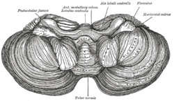 Anterior view of the cerebellum. (Tonsil visible at center right.) | |
 Sagittal section of the cerebellum, near the junction of the vermis with the hemisphere. (Tonsil visible at bottom center.) | |
| Details | |
| Part of | Cerebellum |
| Artery | PICA |
| Identifiers | |
| Latin | tonsilla cerebelli |
| NeuroNames | hier-668 |
| NeuroLex ID | Hemispheric Lobule IX |
| TA | A14.1.07.222 |
| FMA | 83464 |
The cerebellar tonsil is analogous to a rounded lobule on the undersurface of each cerebellar hemisphere, continuous medially with the uvula of the cerebellar vermis and superiorly by the flocculonodular lobe (source: Biology Online) Synonyms include: tonsilla cerebelli, amygdala cerebelli, the latter of which is not to be confused with the cerebral tonsils or amygdala nuclei located deep within the medial temporal lobes of the cerebral cortex. The flocculonodular lobe of the cerebellum which can also be confused for the cerebellar tonsils, is one of three lobes that make up the overall composition of the cerebellum. The cerebellum consists of three anatomical and functional lobes: anterior lobe, posterior lobe, and flocculonodular lobe.
Elongation of the cerebellar tonsils can, due to pressure, lead to this portion of the cerebellum to slip or be pushed through the foramen magnum of the skull resulting is tonsillar herniation. This is a life-threatening condition as it causes increased pressure on the medulla oblongata which contains respiratory and cardiac control centres.
Pathology
A Type I Chiari malformation is a congenital anomaly of the brain in which the cerebellar tonsils are elongated and pushed down through the opening of the base of the skull (see foramen magnum), blocking the flow of cerebrospinal fluid (CSF) as it exits through the medial and lateral apertures of the fourth ventricle. Also called cerebellar tonsillar ectopia, or tonsillar herniation. Although often congenital, Chiari malformation symptoms can also be induced due to physical head trauma, commonly from raised intracranial pressure secondary to a hematoma, or increased dural strain pulling the brain caudally into the foramen magnum. Head trauma increases risk of cerebellar tonsillar ectopia by a factor of 4. Ectopia may be present but asymptomatic until whiplash causes it to become symptomatic.[1]
Additional images
-

Human cerebellum anterior view
-

Human brain midsagittal view
-

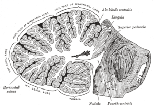
Cerebellum. Inferior surface.
-
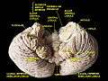
Cerebellum. Inferior surface.
-

Cerebellum. Inferior surface.
References
This article incorporates text in the public domain from the 20th edition of Gray's Anatomy (1918)
- ↑ Freeman, MD (2010). "A case-control study of cerebellar tonsillar ectopia (Chiari) and head/neck trauma (whiplash).". Brain Injury. 24 (7-8): 988–94. doi:10.3109/02699052.2010.490512. PMID 20545453.