White matter
| White matter | |
|---|---|
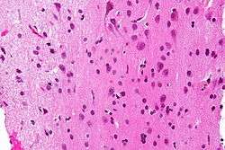 Micrograph showing white matter with its characteristic fine meshwork-like appearance (left of image - lighter shade of pink) and grey matter, with the characteristic neuronal cell bodies (right of image - dark shade of pink). HPS stain. | |
 Human brain right dissected lateral view, showing grey matter (the darker outer parts), and white matter (the inner and prominently whiter parts). | |
| Details | |
| Identifiers | |
| Latin | substantia alba |
| TA | A14.1.00.009 |
| FMA | 83929 |
White matter refers to areas of the central nervous system that are mainly made up of myelinated axons also called tracts.[1] Long thought to be passive tissue, white matter actively affects learning and brain functions, modulating the distribution of action potentials, acting as a relay and coordinating communication between different brain regions.[2]
White matter is named for its relatively light appearance resulting from the lipid content of myelin. However, the tissue of the freshly cut brain appears pinkish white to the naked eye because myelin is composed largely of lipid tissue veined with capillaries. Its white color in prepared specimens is due to its usual preservation in formaldehyde.
Structure
White matter
White matter is composed of bundles of myelinated nerve cell projections (or axons), which connect various gray matter areas (the locations of nerve cell bodies) of the brain to each other, and carry nerve impulses between neurons. Myelin acts as an insulator, increasing the speed of transmission of all nerve signals.[3]
The total number of long range fibers within a cerebral hemisphere is 2% of the total number of cortico-cortical fibers (across cortical areas) and is roughly the same number as those that communicate between the two hemispheres in the brain's largest white tissue structure, the Corpus callosum.[4] Schüz and Braitenberg note "As a rough rule, the number of fibres of a certain range of lengths is inversely proportional to their length."[4]
White matter in nonelderly adults is 1.7–3.6% blood.[5]
Grey matter
The other main component of the brain is grey matter (actually pinkish tan due to blood capillaries), which is composed of neurons. The substantia nigra is a third colored component found in the brain that appears darker due to higher levels of melanin in dopaminergic neurons than its nearby areas. Note that white matter can sometimes appear darker than grey matter on a microscope slide because of the type of stain used. Cerebral- and spinal white matter do not contain dendrites, neural cell bodies, or shorter axons, which can only be found in grey matter.
Location
White matter forms the bulk of the deep parts of the brain and the superficial parts of the spinal cord. Aggregates of gray matter such as the basal ganglia (caudate nucleus, putamen, globus pallidus, subthalamic nucleus, nucleus accumbens) and brain stem nuclei (red nucleus, substantia nigra, cranial nerve nuclei) are spread within the cerebral white matter.
The cerebellum is structured in a similar manner as the cerebrum, with a superficial mantle of cerebellar cortex, deep cerebellar white matter (called the "arbor vitae") and aggregates of grey matter surrounded by deep cerebellar white matter (dentate nucleus, globose nucleus, emboliform nucleus, and fastigial nucleus). The fluid-filled cerebral ventricles (lateral ventricles, third ventricle, cerebral aqueduct, fourth ventricle) are also located deep within the cerebral white matter.
Myelinated axon length
Men have more white matter than females both in volume and in length of myelinated axons. At the age of 20, the total length of myelinated fibers in males is 176,000 km while that of a female is 149,000 km.[6] There is a decline in total length with age of about 10% each decade such that a man at 80 years of age has 97,200 km and a female 82,000 km.[6] Most of this reduction is due to the loss of thinner fibers.[6]
One study found that compared to women, men have approximately 6.5 times the amount of gray matter related to general intelligence; and compared to men, women have nearly 10 times the amount of white matter related to general intelligence. Gray matter represents information processing centers in the brain, and white matter represents the networking of – or connections between – these processing centers.[7]
Function
White matter is the tissue through which messages pass between different areas of gray matter within the central nervous system. The white matter is white because of the fatty substance (myelin) that surrounds the nerve fibers (axons). This myelin is found in almost all long nerve fibers, and acts as an electrical insulation. This is important because it allows the messages to pass quickly from place to place.
Unlike gray matter, which peaks in development in a person's twenties, the white matter continues to develop, and peaks in middle age.[8] This claim has been disputed in recent years, however.
Clinical significance
Multiple sclerosis (MS) is one of the most common diseases which affect white matter. In MS lesions, the myelin shield around the axons has been destroyed by inflammation.
Alcohol use disorders are associated with decrease in white matter volume.[9] Animal studies suggest that alcohol may cause loss of white matter by damaging oligodendrocytes, the glial cell responsible for maintaining myelin.[10]
Changes in white matter known as amyloid plaques are associated with Alzheimer's disease and other neurodegenerative diseases. White matter injuries ("axonal shearing") may be reversible, while gray matter regeneration is less likely. Other changes that commonly occur with age include the development of leukoaraiosis, which is a rarefaction of the white matter that can be caused by a variety of conditions, including loss of myelin, axonal loss, and a breakdown of the blood–brain barrier.
Imaging
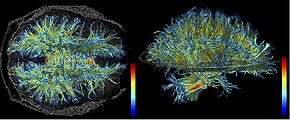
The study of white matter has been advanced with the neuroimaging technique called diffusion tensor imaging where magnetic resonance imaging (MRI) brain scanners are used. As of 2007, more than 700 publications have been published on the subject.[11]
A 2009 paper by Jan Scholz and colleagues[12] used diffusion tensor imaging (DTI) to demonstrate changes in white matter volume as a result of learning a new motor task (e.g. juggling). The study is important as the first paper to correlate motor learning with white matter changes. Previously, many researchers had considered this type of learning to be exclusively mediated by dendrites, which are not present in white matter. The authors suggest that electrical activity in axons may regulate myelination in axons. Or, gross changes in the diameter or packing density of the axon might cause the change.[13] A more recent DTI study by Sampaio-Baptista and colleagues reported changes in white matter with motor learning along with increases in myelination.[14]
References
- ↑ Blumenfeld, Hal (2010). Neuroanatomy through clinical cases (2nd ed.). Sunderland, Mass.: Sinauer Associates. p. 21. ISBN 9780878936137.
Areas of the CNS made up mainly of myelinated axons are called white matter.
- ↑ Douglas Fields, R. (2008). "White Matter Matters". Scientific American. 298 (3): 54–61. Bibcode:2008SciAm.298c..54D. doi:10.1038/scientificamerican0308-54.
- ↑ Klein, S.B., & Thorne, B.M. Biological Psychology. Worth Publishers: New York. 2007.
- 1 2 Schüz, Almut; Braitenberg, Valentino (2002). "The human cortical white matter: Quantitative aspects of cortico-cortical long-range connectivity". In Schüz, Almut; Braitenberg, Valentino. Cortical Areas: Unity and Diversity, Conceptual Advances in Brain Research. Taylor and Francis. pp. 377–86. ISBN 978-0-415-27723-5.
- ↑ Leenders, K. L.; Perani, D.; Lammertsma, A. A.; Heather, J. D.; Buckingham, P.; Jones, T.; Healy, M. J. R.; Gibbs, J. M.; Wise, R. J. S.; Hatazawa, J.; Herold, S.; Beaney, R. P.; Brooks, D. J.; Spinks, T.; Rhodes, C.; Frackowiak, R. S. J. (1990). "Cerebral Blood Flow, Blood Volume and Oxygen Utilization". Brain. 113: 27–47. doi:10.1093/brain/113.1.27. PMID 2302536.
- 1 2 3 Marner, Lisbeth; Nyengaard, Jens R.; Tang, Yong; Pakkenberg, Bente (2003). "Marked loss of myelinated nerve fibers in the human brain with age". The Journal of Comparative Neurology. 462 (2): 144–52. doi:10.1002/cne.10714. PMID 12794739.
- ↑ University Of California, Irvine. "Intelligence In Men And Women Is A Gray And White Matter." ScienceDaily. ScienceDaily, 22 January 2005. <www.sciencedaily.com/releases/2005/01/050121100142.htm>.
- ↑ Sowell, Elizabeth R.; Peterson, Bradley S.; Thompson, Paul M.; Welcome, Suzanne E.; Henkenius, Amy L.; Toga, Arthur W. (2003). "Mapping cortical change across the human life span". Nature Neuroscience. 6 (3): 309–15. doi:10.1038/nn1008. PMID 12548289.
- ↑ Monnig, Mollie A.; Tonigan, J. Scott; Yeo, Ronald A.; Thoma, Robert J.; McCrady, Barbara S. (2013). "White matter volume in alcohol use disorders: A meta-analysis". Addiction Biology. 18 (3): 581–92. doi:10.1111/j.1369-1600.2012.00441.x. PMC 3390447
 . PMID 22458455.
 - ↑ Alfonso-Loeches, Silvia; Pascual, Maria; Gómez-Pinedo, Ulises; Pascual-Lucas, Maya; Renau-Piqueras, Jaime; Guerri, Consuelo (2012). "Toll-like receptor 4 participates in the myelin disruptions associated with chronic alcohol abuse". Glia. 60 (6): 948–64. doi:10.1002/glia.22327. PMID 22431236.
- ↑ Assaf, Yaniv; Pasternak, Ofer (2007). "Diffusion Tensor Imaging (DTI)-based White Matter Mapping in Brain Research: A Review". Journal of Molecular Neuroscience. 34 (1): 51–61. doi:10.1007/s12031-007-0029-0. PMID 18157658.
- ↑ Scholz, Jan; Klein, Miriam C; Behrens, Timothy E J; Johansen-Berg, Heidi (2009). "Training induces changes in white-matter architecture". Nature Neuroscience. 12 (11): 1370–1. doi:10.1038/nn.2412. PMC 2770457. PMID 19820707.
- ↑ "White Matter Matters". Dolan DNA Learning Center. Archived from the original on 2009-11-12. Retrieved 2009-10-19.
- ↑ Sampaio-Baptista, C.; Khrapitchev, A. A.; Foxley, S.; Schlagheck, T.; Scholz, J.; Jbabdi, S.; Deluca, G. C.; Miller, K. L.; Taylor, A.; Thomas, N.; Kleim, J.; Sibson, N. R.; Bannerman, D.; Johansen-Berg, H. (2013). "Motor Skill Learning Induces Changes in White Matter Microstructure and Myelination". Journal of Neuroscience. 33 (50): 19499–503. doi:10.1523/JNEUROSCI.3048-13.2013. PMC 3858622. PMID 24336716.
External links
| Wikimedia Commons has media related to White matter. |
- White Matter Atlas
- WebMD (2009). "white matter". Webster's New World Medical Dictionary (3rd ed.). Houghton Mifflin Harcourt. p. 456. ISBN 978-0-544-18897-6.