Corneal limbus
For other uses, see Limbus.
| Corneal limbus | |
|---|---|
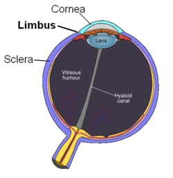
 Schematic diagram of the human eye | |
| Details | |
| Identifiers | |
| Latin | Limbus corneae |
| TA | A15.2.02.014 |
| FMA | 58342 |
The corneal limbus is the border of the cornea and the sclera (the white of the eye). The limbus is a common site for the occurrence of corneal epithelial neoplasm. The limbus contains radially-oriented fibrovascular ridges known as the palisades of Vogt that may harbour a stem cell population.[1] The palisades of Vogt are more common in the superior and inferior quadrants around the eye.[2] Aniridia, a developmental anomaly of the iris, disrupts the normal barrier of the cornea to the conjunctival epithelial cells at the limbus.
Additional images
-
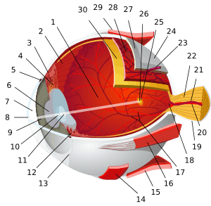
Extrinsic eye muscle. Nerves of orbita. Deep dissection.
See also
References
- ↑ Thomas PB, Liu YH, Zhuang FF, Selvam S, Song SW, Smith RE, Trousdale MD, Yiu SC (2007). "Identification of Notch-1 expression in the limbal basal epithelium". Mol. Vis. 13: 337–44. PMC 2633467
 . PMID 17392684.
 - ↑ Goldberg MF, Bron AJ (1982). "Limbal palisades of Vogt". Transactions of the American Ophthalmological Society. 80: 155–71. PMC 1312261
 . PMID 7182957.
External links
- Atlas image: eye_1 at the University of Michigan Health System - "Sagittal Section Through the Eyeball"
- http://www.vetmed.ucdavis.edu/courses/vet_eyes/images/s_4021_2.jpg
This article is issued from Wikipedia - version of the 11/23/2016. The text is available under the Creative Commons Attribution/Share Alike but additional terms may apply for the media files.

