Foveola
| Foveola | |
|---|---|
| Details | |
| Identifiers | |
| Latin | foveola |
| TA | A15.2.04.023 |
| FMA | 77666 |
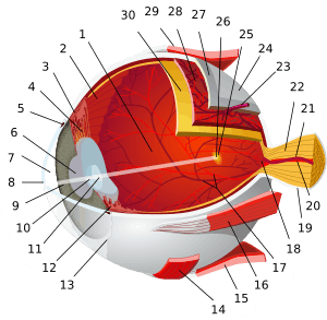
The foveola is located within a region called the macula, a yellowish, cone photo receptor filled portion of the human retina. The foveola is approximately 0.35 mm in diameter and lies in the center of the fovea and contains only cone cells, and a cone-shaped zone of Müller cells.[1] In this region the cone receptors are found to be longer, slimmer and more densely packed than anywhere else in the retina, thus allowing that region to have the potential to have the highest visual acuity in the eye.
Notes
- ↑ Gass, JD (Jun 1999). "Müller cell cone, an overlooked part of the anatomy of the fovea centralis: hypotheses concerning its role in the pathogenesis of macular hole and foveomacular retinoschisis.". Archives of Ophthalmology. 117 (6): 821–3. doi:10.1001/archopht.117.6.821. PMID 10369597.
This article is issued from Wikipedia - version of the 8/4/2016. The text is available under the Creative Commons Attribution/Share Alike but additional terms may apply for the media files.

