Cribriform plate
| Cribriform plate | |
|---|---|
 Ethmoid bone from above. | |
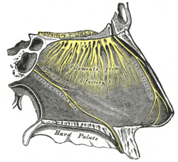 Nerves of septum of nose. Right side. | |
| Details | |
| Identifiers | |
| Latin | lamina cribrosa ossis ethmoidalis |
| TA | A02.1.07.002 |
| FMA | 52890 |
The cribriform plate of the ethmoid bone (horizontal lamina or lamina cribrosa) is received into the ethmoidal notch of the frontal bone and roofs in the nasal cavities.
Structure
Projecting upward from the middle line of this plate is a thick, smooth, triangular process, the crista galli, so called from its resemblance to a rooster's comb.
The long thin posterior border of the crista galli serves for the attachment of the falx cerebri.
Its anterior border, short and thick, articulates with the frontal bone, and presents two small projecting alae (wings), which are received into corresponding depressions in the frontal bone and complete the foramen cecum.
Its sides are smooth, and sometimes bulging from presence of a small air sinus in the interior.
On either side of the crista galli, the cribriform plate is narrow and deeply grooved; it supports the olfactory bulb and is perforated by foramina for the passage of the olfactory nerves. The foramina in the middle of the groove are small and transmit the nerves to the roof of the nasal cavity; those at the medial and lateral parts of the groove are larger—the former transmit the nerves to the upper part of the nasal septum, the latter those to the superior nasal concha.
At the front part of the cribriform plate, on either side of the crista galli, is a small fissure that is occupied by a process of dura mater.
Lateral to this fissure is a notch or foramen which transmits the nasociliary nerve; from this notch a groove extends backward to the anterior ethmoidal foramen.
Clinical significance
A fractured cribriform plate can result in leaking of cerebrospinal fluid into the nose and loss of sense of smell. The tiny apertures of the plate transmitting the olfactory nerve become the route of ascent for a pathogen, Naegleria fowleri. The pathogen tends to destroy the olfactory bulb and the adjacent inferior surface of the frontal lobe of the brain. This surface initially becomes the site of proliferation of the trophozoites of Naegleria fowleri and their subsequent spread to the rest of the brain and CSF. Because of its initial involvement and trophozoite presence in early phases of Naegleria fowleri infection, flushing of this region with saline using a device, to obtain Naegelria fowleri for diagnostic PCR and microscopic viewing has been proposed for patients affected by Primary Amoebic Meningoencephalitis (PAM), by (Baig AM., et al) in a recent publication.[1] The latter scientists have suggested the same route to administer drugs at an early phase of infection by using a " Transcribrial Route Device"[2] that has been proposed to kill this pathogen at a place of its maximum proliferation. A recent Australian study as shown that bacterium causing the tropical disease melioidosis, Burkholderia pseudomallei can also invade the brain via the olfactory nerve within 24 h by transversing the cribriform plate.[3]
Keros Classification
The Keros classification is a method of classifying the depth of the olfactory fossa.
The depth of the olfactory fossa is determined by the height of the lateral lamella of the cribriform plate. Keros in 1962, classified the depth into three categories.
- type 1: has a depth of 1–3 mm (26.3% of population)
- type 2: has a depth of 4-7mm (73.3% of population)
- type 3: has a depth of 8-16mm (0.5% of population)
History
From Latin cribrum ("sieve") + -form.
Additional images
-
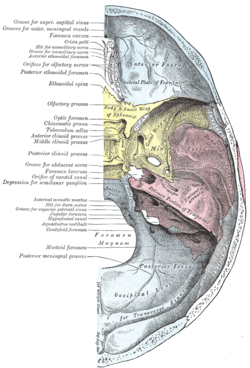
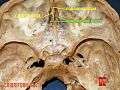
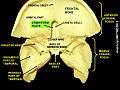
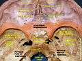
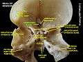
Base of the skull. Upper surface.
-
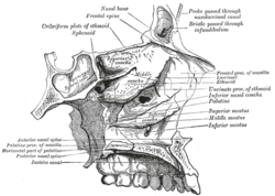
Roof, floor, and lateral wall of left nasal cavity.
-

Nerves of the orbit. Seen from above.
-
Cribriform plate
-
Cribriform plate
-
Cribriform plate
-
Cribriform plate
References
This article incorporates text in the public domain from the 20th edition of Gray's Anatomy (1918)
- ↑ Abdul Mannan Baig, Naveed Ahmed Khan, (2014). Tackling infection owing to brain-eating amoeba. Acta Tropica 11/2014; doi: 10.1016/j.actatropica.2014.11.004
- ↑ Abdul Mannan Baig, Naveed Ahmed Khan,. , (2014) Novel chemotherapeutic strategies in the management of primary amoebic meningoencephalitis due to Naegleria fowleri. CNS Neurosciences & Therapeutics (2014). 01/2014; doi:10.1111/cns.12225
- ↑ St John, JA; Ekberg, JA; Dando, SJ; Meedeniya, AC; Horton, RE; Batzloff, M; Owen, SJ; Holt, S; Peak, IR; Ulett, GC; Mackay-Sim, A; Beacham, IR (Apr 2014). "Burkholderia pseudomallei penetrates the brain via destruction of the olfactory and trigeminal nerves: implications for the pathogenesis of neurological melioidosis". MBio. 5 (2): e00025. doi:10.1128/mBio.00025-14. PMC 3993850
 . PMID 24736221.
External links
- Cross section image: skull/x-front - Plastination Laboratory at the Medical University of Vienna