External occipital protuberance
Near the middle of the squamous part of occipital bone is the external occipital protuberance, the highest point of which is referred to as the inion. The inion is the most prominent projection of the protuberance which is located at the posterioinferior (lower rear) part of the human skull. The nuchal ligament and trapezius muscle attach to it.
The inion (ἰνίον, iníon, Greek for the occipital bone) is used as a landmark in the 10-20 system in EEG recording.
Extending laterally from it on either side is the superior nuchal line, and above it is the faintly marked highest nuchal line.
A study of 16th-century Anatolian remains showed that the external occipital protuberance statistically tends to be less pronounced in female remains.[1]
Additional images
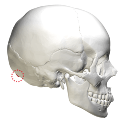
| Position of external occipital protuberance (shown in red). Animation. |
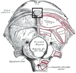
| Occipital bone. Position of external occipital protuberance shown in red. |
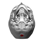
| Human skull seen from below. Position of external occipital protuberance shown in red. |
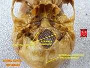
| Human skull seen from below. External occipital protuberance labelled at the bottom. |
| Occipital bone replica of Homo erectus (400,000 years old) seen from behind. |
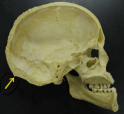
| Midsaggital section of a human skull. Inion indicated by yellow arrow. |
|
See also
References
This article incorporates text in the public domain from the 20th edition of Gray's Anatomy (1918)
External links