Lateral plate mesoderm
| Lateral plate mesoderm | |
|---|---|
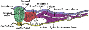 Transverse section of a chick embryo of forty-five hours’ incubation. * Chordamesoderm: yellow, at notochord. * Paraxial mesoderm: red, at somite. * Intermediate mesoderm: purple, near Wolffian duct. * Lateral plate mesoderm: purple, near "Somatic mesoderm" and "Splanchic mesoderm". | |
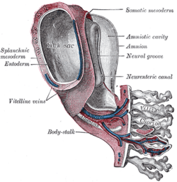 Model of human embryo 1.3 mm. long.. (Splanchic mesoderm labeled at left, somatic mesoderm at top right.) | |
| Details | |
| Carnegie stage | 9 |
| Precursor | mesoderm |
| Gives rise to | somatopleure, splanchnopleure |
| Identifiers | |
| Latin | mesoderma laminae lateralis |
| Code | TE E5.0.3.0.0.0.2 |
Lateral plate mesoderm is a type of mesoderm that is found at the periphery of the embryo.
Division into layers
It will split into two layers, the somatic layer/mesoderm and the splanchnic layer/mesoderm
- The somatopleuric layer forms the future body wall.
- The splanchnopleuric layer forms the circulatory system.
Spaces within the lateral plate are enclosed and forms the intraembryonic coelom.
It is formed by the secretion of BMP-4 by the ectoderm.[1]
Serosal mesoderms
Lateral plate mesoderm gives rise to the serosal mesoderms.[2]
- forms a ventral layer associated with endoderm, the splanchnopleuric mesoderm. This forms the viscera and heart
- forms a dorsal layer associated with ectoderm, the somatopleuric mesoderm. This forms the body wall lining and dermis.
- Abdominal portion becomes contained in dorsal mesentery, part of the serosal mesoderm.
- When the two layers form, a cardiogenic plate is visible. Later, this will form the myocardial primordium, which will contribute to the tubular heart.
Cavities
In the 4th week the coelom divides into pericardial, pleural and peritoneal cavities.[2]
- First partition: is the septum transversum.
- This will be translocated later into the diaphragm and ventral mesentery.
- Divides the coelom into primitive pericardial and peritoneal cavities
- Pleuropericardial folds appear on the lateral wall of primitive pericardial cavity, which will eventually cause a partition to form the pericardial and pleural cavities.
- Communication between these partitions formed by the pericardioperitoneal canals. However, pleuroperitoneal membranes will grow to fuse with the septum transversum to close off these canals.
- At day 22, lung buds form, remaining ensheathed in a splanchnopleuric mesoderm
Limb Development
Cells from the lateral plate mesoderm and the myotome migrate to the limb field and proliferate to create the limb bud. The lateral plate cells produce the cartilaginous and skeletal portions of the limb while the myotome cells produce the muscle components. The lateral plate mesodermal cells secrete a fibroblast growth factor (FGF7 and FGF10, presumably) to induce the overlying ectoderm to form an important organizing structure called the apical ectodermal ridge (AER).The AER reciprocatively secretes FGF8 and FGF4 which maintains the FGF10 signal and induces proliferation in the mesoderm. The position of FGF10 expression is regulated by Wnt8c in the hindlimb and Wnt2b in the forelimb. The forelimb and the hindlimb are specified by their position along the anterior/posterior axis and possibly by two T-box containing transcription factors: Tbx5 and Tbx4, respectively.
See also
- Limb development for more information
- Serous membrane
References
This article incorporates text in the public domain from the 20th edition of Gray's Anatomy (1918)
- ↑ Tonegawa A, Funayama N, Ueno N, Takahashi Y (1997). "Mesodermal subdivision along the mediolateral axis in chicken controlled by different concentrations of BMP-4". Development. 124 (10): 1975–84. PMID 9169844.
- 1 2 Larsen, William J. (1998). Essentials of human embryology. Edinburgh: Churchill Livingstone. ISBN 0-443-07514-X.
External links
- Swiss embryology (from UL, UB, and UF) hdisqueembry/triderm08