Mediastinum
| Mediastinum | |
|---|---|
 Mediastinum. The division between superior and inferior is at the sternal angle. | |
 Mediastinum anatomy | |
| Details | |
| Identifiers | |
| Latin | mediastinus |
| TA | A07.1.02.101 |
| FMA | 9826 |
The mediastinum (from Medieval Latin mediastinus, "midway"[1]) is the central compartment of the thoracic cavity surrounded by loose connective tissue, as an undelineated region that contains a group of structures within the thorax. The mediastinum contains the heart and its vessels, the esophagus, trachea, phrenic and cardiac nerves, the thoracic duct, thymus and lymph nodes of the central chest.
Structure
The mediastinum lies within the thorax and is enclosed on the right and left by pleurae. It is surrounded by the chest wall in front, the lungs to the sides and the spine at the back. It extends from the sternum in front to the vertebral column behind, and contains all the organs of the thorax except the lungs. It is continuous with the loose connective tissue of the neck.
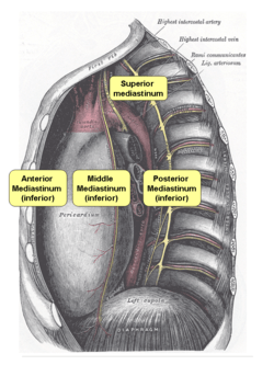
The mediastinum can be divided into an upper (or superior) and lower (or inferior) part:
- The superior mediastinum starts at the superior thoracic aperture and ends at the thoracic plane.
- The thoracic plane separates the superior and inferior mediastinum. It is a plane at the level of the sternal angle , and the intervertebral disc of T4–T5.[2][3][4]
- The inferior mediastinum from this level to the diaphragm. This lower part is subdivided into three regions, all relative to the pericardium - the anterior mediastinum being in front of the pericardium, the middle mediastinum contains the pericardium and its contents, and the posterior mediastinum being behind the pericardium.
Anatomists, surgeons, and clinical radiologists compartmentalize the mediastinum differently. For instance, in the radiological scheme of Felson, there are only three compartments (anterior, middle, and posterior), and the heart is part of the anterior mediastinum.[5]
Superior mediastinum
The superior mediastinum is bounded:
- superiorly by the thoracic inlet, the upper opening of the thorax;
- inferiorly by the transverse thoracic plane;
- laterally by the pleurae;
- anteriorly by the manubrium of the sternum;
- posteriorly by the first four thoracic vertebral bodies.
| Contents |
|---|
|
Thoracic plane
- The thoracic plane separates the superior and inferior mediastinum. It is a plane at the level of the sternal angle, and the intervertebral disc of T4–T5.[6][7][8]
A number of structures occur at the level of the thoracic plane, which divides the superior and inferior mediastinum:
| Structures at the level of the thoracic plane |
|---|
|
Inferior mediastinum
- Anterior mediastinum
Is bounded:
- laterally by the pleurae;
- posteriorly by the pericardium;
- anteriorly by the sternum, the left transversus thoracis and the fifth, sixth, and seventh left costal cartilages.
| Contents |
|---|
|
- Middle mediastinum
Bounded: pericardial sac – It contains the vital organs and is classified into the serous and fibrous pericardium.
| Contents |
|---|
|
- Posterior mediastinum
Is bounded:
- anteriorly by the pericardium (in front of);
- inferiorly by the thoracic surface of the diaphragm (below);
- superiorly by the transverse thoracic plane;
- posteriorally by the bodies of the vertebral column from the lower border of the fifth to the twelfth thoracic vertebra (behind);
- laterally by the mediastinal pleura (on either side).
| Contents |
|---|
|
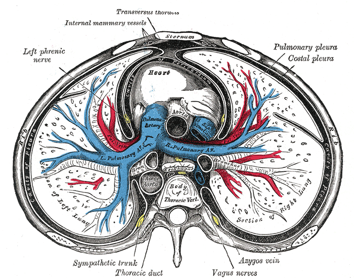 A transverse section of the thorax, showing the contents of the middle and the posterior mediastinum.
Clinical significance
The mediastinum is frequently the site of involvement of various tumors:
- Anterior mediastinum: substernal thyroid goiters, lymphoma, thymoma, and teratoma.
- Middle mediastinum: lymphadenopathy, metastatic disease such as from small cell carcinoma from the lung.
- Posterior mediastinum: Neurogenic tumors, either from the nerve sheath (mostly benign) or elsewhere (mostly malignant).
Mediastinitis is inflammation of the tissues in the mediastinum, usually bacterial and due to rupture of organs in the mediastinum. As the infection can progress very quickly, this is a serious condition.
Pneumomediastinum is the presence of air in the mediastinum, which in some cases can lead to pneumothorax, pneumoperitoneum, and pneumopericardium if left untreated. However, that does not always occur and sometimes those conditions are actually the cause, not the result, of pneumomediastinum. These conditions frequently accompany Boerhaave's syndrome, or spontaneous esophageal rupture.
There are many diseases that can present with a widened mediastinum (usually found via a chest x-ray). The most common ones are aortic unfolding, traumatic aortic rupture, thoracic aortic aneurysm, and traumatic thoracic vertebral fracture. A widened mediastinum is a classic but rare hallmark sign of anthrax infection.
See also
- Anthrax
- Mediastinum testis (unrelated structure in the scrotum)
- Mediastinal germ cell tumor
- Mediastinitis
- Mediastinal tumor
- Widened mediastinum
References
This article incorporates text in the public domain from the 20th edition of Gray's Anatomy (1918)
- ↑ Mediastinum dictionary definition
- ↑ Thoracic Wall, Pleura, and Pericardium - Dissector Answers
- ↑ Untitled Document
- ↑ UAMS Department of Anatomy - Topographical Anatomy - Thorax
- ↑ Goodman, Lawrence. Felson's Principles of Chest Roentgenology.
- ↑ Thoracic Wall, Pleura, and Pericardium - Dissector Answers
- ↑ Untitled Document
- ↑ UAMS Department of Anatomy - Topographical Anatomy - Thorax
- ↑ Arai et al. Radiographic landmarks of the upper margin of the superior vena cava (SVC) in children Canadian Journal of Anesthesia 49 (Supplement 1): 32
- ↑ Viscera of the Thorax UAMS Department of Anatomy
External links
| Look up mediastinum in Wiktionary, the free dictionary. |
- 1691353147 at GPnotebook
- Anatomy figure: 21:01-03 at Human Anatomy Online, SUNY Downstate Medical Center – "Divisions of the mediastinum."
- Anatomy figure: 21:02-03 at Human Anatomy Online, SUNY Downstate Medical Center – "The anatomical divisions of the inferior mediastinum."
- thoraxlesson3 at The Anatomy Lesson by Wesley Norman (Georgetown University) – "Subdivisions of the Thoracic Cavity"