Microrheology
Microrheology[1] is a technique used to measure the rheological properties of a medium, such as microviscosity, via the measurement of the trajectory of a flow tracer (a micrometre-sized particle). It is a new way of doing rheology, traditionally done using a rheometer. There are two types of microrheology: passive microrheology and active microrheology. Passive microrheology uses inherent thermal energy to move the tracers, whereas active microrheology uses externally applied forces, such as from a magnetic field or an optical tweezer, to do so. Microrheology can be further differentiated into 1- and 2-particle methods.[2] [3]
Passive microrheology
Passive microrheology uses the thermal energy (kT) to move the tracers, although recent evidence suggest that active random forces inside cells may instead move the tracers in a diffusive-like manner.[4] The trajectories of the tracers are measured optically either by microscopy or by diffusing-wave spectroscopy (DWS). From the mean squared displacement with respect to time (noted MSD or <Δr2> ), one can calculate the visco-elastic moduli G′(ω) and G″(ω) using the generalized Stokes–Einstein relation (GSER). Here is a view of the trajectory of a particle of micrometer size.
-
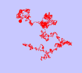
Typical trajectory of a Brownian particle (simulation)
-
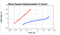
Two examples of MSD: one for a purely viscous fluid (free diffusion) and one for a viscolelastic fluid (trapped by elastic network)
-
Animation of a particle in a polymer-like network
Observing the MSD for a wide range of time scales gives information on the microstructure of the medium where are diffusing the tracers. If the tracers are having a free diffusion, one can deduce that the medium is purely viscous. If the tracers are having a sub-diffusive mean trajectory, it indicates that the medium presents some viscoelastic properties. For example, in a polymer network, the tracer may be trapped. The excursion δ of the tracer is related to the elastic modulus G′ with the relation G′ = kBT/(6πaδ2).[5]
Microrheology is another way to do linear rheology. Since the force involved is very weak (order of 10−15 N), microrheology is guaranteed to be in the so-called linear region of the strain/stress relationship. It is also able to measure very small volumes (biological cell).
Given the complex viscoelastic modulus with G′(ω) the elastic (conservative) part and G″(ω) the viscous (dissipative) part and ω=2πf the pulsation. The GSER is as follows:


with
- : Laplace transform of G
- kB: Boltzmann constant
- T: temperature in kelvins
- s: the Laplace frequency
- a: the radius of the tracer
- : the Laplace transform of the mean squared displacement


A related method of passive microrheology involves the tracking positions of a particle at a high frequency, often with a quadrant photodiode.[6] From the position, , the power spectrum, can be found, and then related to the real and imaginary parts of the response function, .[7] The response function leads directly to a calculation of the complex shear modulus, via:


Active microrheology
Active microrheology may use a magnetic field [8][9][10][11][12] or optical tweezers[13][14][15] to apply a force on the tracer and then find the stress/strain relation. More recently, it has been developed into Force spectrum microscopy to measure contributions of random active motor proteins to diffusive motion in the cytoskeleton.[4]
References
- ↑ Mason, Thomas G. & Weitz, David A. (1995). "Optical Measurements of Frequency-Dependent Linear Viscoelastic Moduli of Complex Fluids". Physical Review Letters. 74: 7. Bibcode:1995PhRvL..74.1250M. doi:10.1103/physrevlett.74.1250.
- ↑ Crocker, John C.; Valentine, M. T.; Weeks, Eric R.; Gisler, T.; et al. (2000). "Two-Point Microrheology of Inhomogeneous Soft Materials". Physical Review Letters. 85 (4): 888–891. Bibcode:2000PhRvL..85..888C. doi:10.1103/PhysRevLett.85.888.
- ↑ Levine, Alex J. & Lubensky, T. C. (2000). "One- and Two-Particle Microrheology". Physical Review Letters. 85 (8): 1774–1777. arXiv:cond-mat/0004103
 . Bibcode:2000PhRvL..85.1774L. doi:10.1103/PhysRevLett.85.1774.
 - 1 2 Guo, Ming; et al. (2014). "Probing the Stochastic, Motor-Driven Properties of the Cytoplasm Using Force Spectrum Microscopy". Cell (158): 822–832. doi:10.1016/j.cell.2014.06.051.
- ↑ Bellour, M.; Skouri, M.; Munch, J.-P.; Hébraud, P. (2002). "Brownian motion of particles embedded in a solution of giant micelles". European Physical Journal E. 8: 431–436. Bibcode:2002EPJE....8..431B. doi:10.1140/epje/i2002-10026-0.
- ↑ Schnurr, B.; Gittes, F.; MacKintosh, F. C. & Schmidt, C. F. (1997). "Determining Microscopic Viscoelasticity in Flexible and Semiflexible Polymer Networks from Thermal Fluctuations". Macromolecules. 30 (25): 7781–7792. arXiv:cond-mat/9709231. Bibcode:1997MaMol..30.7781S. doi:10.1021/ma970555n.
- ↑ Gittes, F.; Schnurr, B.; Olmsted, P. D.; MacKintosh, F. C.; et al. (1997). "Determining Microscopic Viscoelasticity in Flexible and Semiflexible Polymer Networks from Thermal Fluctuations". Physical Review Letters. 79 (17): 3286–3289. arXiv:cond-mat/9709228. Bibcode:1997PhRvL..79.3286G. doi:10.1103/PhysRevLett.79.3286.
- ↑ A.R. Bausch; et al. (1999). "Measurement of local viscoelasticity and forces in living cells by magnetic tweezers". Biophysical Journal. 76 (1 Pt 1): 573–9. Bibcode:1999BpJ....76..573B. doi:10.1016/S0006-3495(99)77225-5. PMC 1302547. PMID 9876170.
- ↑ K.S. Zaner & P.A. Valberg (1989). "Viscoelasticity of F-actin measured with magnetic microparticles". Journal of Cell Biology. 109 (5): 2233–43. doi:10.1083/jcb.109.5.2233. PMC 2115855. PMID 2808527.
- ↑ F.Ziemann; J. Radler & E. Sackmann (1994). "Local measurements of viscoelastic moduli of entangled actin networks using an oscillating magnetic bead micro-rheometer". Biophysical Journal. 66 (6): 2210–6. Bibcode:1994BpJ....66.2210Z. doi:10.1016/S0006-3495(94)81017-3. PMC 1275947. PMID 8075354.
- ↑ F.G. Schmidt; F. Ziemann & E. Sackmann (1996). "Shear field mapping in actin networks by using magnetic tweezers". European Biophysics Journal. 24: 348. doi:10.1007/bf00180376.
- ↑ F. Amblard; et al. (1996). "Subdiffusion and Anomalous Local Viscoelasticity in Actin Networks". Physical Review Letters. 77 (21): 4470–4473. Bibcode:1996PhRvL..77.4470A. doi:10.1103/PhysRevLett.77.4470. PMID 10062546.
- ↑ E. Helfer; et al. (2000). "Microrheology of Biopolymer-Membrane Complexes". Physical Review Letters. 85 (2): 457–60. Bibcode:2000PhRvL..85..457H. doi:10.1103/PhysRevLett.85.457. PMID 10991307.
- ↑ Manlio Tassieri; et al. (2012). "Microrheology with optical tweezers: data analysis". New Journal of Physics. 14 (11): 115032. Bibcode:2012NJPh...14k5032T. doi:10.1088/1367-2630/14/11/115032.
- ↑ David Engström; Michael C.M. Varney; Martin Persson; Rahul P. Trivedi; et al. (2012). "Unconventional structure-assisted optical manipulation of high-index nanowires in liquid crystals". Optics Express. 20 (7): 7741–7748. Bibcode:2012OExpr..20.7741E. doi:10.1364/OE.20.007741.
External links
- Harvard Weitz Lab page
- Review of microrheology in optical tweezers
- Review on microrheology
- Illustrated description of microrheology and a microrheology analysis, with movies
- Microrheology Overview with Animations