Muscina
| Muscina | |
|---|---|
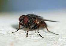 | |
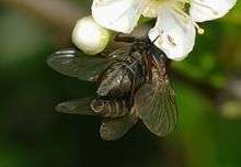
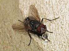
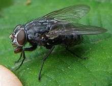
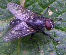
| Adult Muscina stabulans | |
| Scientific classification | |
| Kingdom: | Animalia |
| Phylum: | Arthropoda |
| Class: | Insecta |
| Order: | Diptera |
| Family: | Muscidae |
| Subfamily: | Azeliinae[1] |
| Tribe: | Reinwardtiini |
| Genus: | Muscina[2] Robineau-Desvoidy, 1830 |
| Type species | |
| M. stabulans Fallén, 1817[3] | |
Muscina is a genus of flies that belongs to the family Muscidae, currently consisting of 27 species.[2] They are worldwide in distribution and are frequently found in livestock facilities and outside restrooms. The most common species are M. stabulans (the most widely studied species), M. levida, and M. prolapsa. Muscina flies commonly breed in manure and defecate on food, which has been linked to the spread of some disease and illnesses.[4][5] The occurrence of Muscina larvae on dead bodies has led to their regular use in forensic investigations, as they may be used to estimate the time of death.[6] Research have shown the prevalence of certain species of Muscina flies as vectors of diseases such as poliomyelitis.[7]
Characteristics
Muscina species are characterized by a retractable proboscis, sponging or sucking mouthparts, and a pale tip on the scutellum.[8][4] The species M. stabulans and M. levida are larger than the housefly, and have moderately curved fourth veins with the latter also having a black palpi. The species M. levida has legs that are entirely black. M. pascuorum flies have a red palpi, a strongly curved fourth vein that ends in or before the wing tip, and are generally larger than M. levida.[8]
Muscina stabulans
The species M. stabulans, more commonly known as the false stable fly, has partially reddish-brown legs, four characteristic dark stripes along the thorax region, and a pale spot above the thorax. These flies average 8 millimeters (0.3 inches) in length.[4] The abdomen of the false stable fly is either entirely black, or black with red sides. Its head ranges in color from a dark-grey to a whitish hue.[5] Circular spiracular plates can be found separated by about one plate’s width in the posterior area.[9]
The M. stabulans species were found in a study determining synanthropy (ecologically associated with humans) with the adult Muscidae species collected in urban, rural and natural environments in Valdivia, Chile. According to the study, M. stabulans are mostly found in rural and natural neighborhoods, and rarely inhabit cities.[10] The distribution of Muscina species tends to be spread throughout the United States. For the most part, Muscina stabulan is active year round, but fly activity tends to peak in the summer months, when the number of generation cycles also peak.
The habitat of the M. stabulans is similar to that of the house fly, Musca domestica. M. stabulans have been spotted in animal housing, such as poultry houses, as well as in the mucosal linings of mammalian intestines. M. stabulan can be found on carrions in the decomposition stages, but they exhibit a strong preference for the later stages of decomposition. They are able to reach a buried body in shallow ground through several inches of dirt.[11]
Life cycle
Muscina species undergo the same transformations throughout the life cycle as similar species and families in the order Diptera. Flies in the Dipteran order undergo what is known as holometabolous transformation, a type of metabolic transformation in which an insect starts out as an egg, undergoes larval stages, and then pupates before reaching full adulthood. The adult insect is referred to as an imago.[12]
Eggs and larvae
Flies are completely wingless at the beginning of development. In normal fly production there can be as many as 250 eggs laid by a mature female.[13] The adult female keeps the eggs inside the abdomen until all of the eggs are produced and then lays the eggs through an ovipositor located on the hind end of the abdomen.
These eggs are very small in size. Female adult flies usually choose decaying matter as sites to lay the eggs. These nutrient-rich areas are ideal for the growth and development of the hatching larvae. In contrast to adult flies, the larvae do not have a definite head. Instead, there are two grasping hooks that they use to cut and tear food.[14] The larvae of Muscina have 11 segments. At the posterior end of the larvae are a set of spiracles. The spiracles of Muscina have spiracular slits that are not straight and exhibit some form of curvature.[15] Larvae use the spiracles to breathe. The spiracles have a number of slits that can be used to determine what instar, or larval stage, the larvae is in; for example, one slit means first instar, two slits means second instar, and three slits means third instar.[14] It has been shown that the environmental temperature has a strong influence on larval development: increasing temperature directly influences the amount of time that the larvae need to complete development.[16]
Pupae
After the larval phase, the Muscina larvae pass through a pupal stage. In this stage, there are many transformations that occur, such as the formation of legs, head, and wings. Simultaneously, a protective layer builds up and forms a cocoon, which aids in protection of the vital organs of the fly. M. levida is a species in this genus that does not form a cocoon. The duration of the pupal stage also varies depending on the temperature of the surrounding environment.[16]
Adults
After sufficient time for pupal development has elapsed, the fly will be able to break out of its hard pupa shell, and the fly’s wings begin to spread. Although completely formed in the pupa stage, the adult’s wings do not reach its full size until outside the pupa covering. Through the use of blood vessels inside the wings, the fly is able to expand to full width and length and complete its life cycle.[14]
Medical importance
Some insects have been shown to be potential carriers of pathogenic agents that can cause diseases. Mosquitoes and ticks as well as certain species of Muscina flies have been revealed to be possible vectors. M. stabulans, along with almost two dozen other species of flies have been named the “disease-causing flies.”[17] Species of flies such as M. stabulans can spread bacterial and viral pathogens via transfer from its feet or mouthparts. Adult female flies tend to lay eggs in decaying material such as food or dead organisms and fresh fecal material. The fecal material houses a vast number of pathogenic bacteria, viruses, protozoan and other disease-causing agents. Most of the bacteria and viruses are not introduced from the fecal material to the fly when in the egg or larvae form; rather, the transfer occurs in the transition of a young fly to adulthood. Fecal particles attach to the fly’s outer body as it emerges from the larvae. Transfer of bacteria occurs when the fly takes off and lands on an open wound or food material. Physical contact flakes the pathogen off the fly's body and causes contamination. The spread of a pathogen by means of a fly’s outer body, such as its feet, to the host, is referred to as mechanical transmission. It is possible to determine the identify pathogen carried by identifying the species of fly. In these instances, food sanitation is an important preventive measure to ensure food safety.[18] Moreover, a study of flies including M. stabulans and M. levida has shown that fly incidence peaked about 4–5 months before the occurrence of a poliomyelitis epidemic. This time period matches the time it takes for the infective agent to incubate in a human plus the extra time necessary for the fly to acquire and incubate the virus in its body.[7]
Forensic importance
M. stabulans and M. levida belong to the ecological group of the filth fly.[9] Muscina flies are attracted to decaying organic matter, and are commonly found on corpses, urine, and feces. Muscina flies are useful in determining post-mortem intervals. The presence of Muscina larvae in diapers and on genitalia can indicate a timeline for the period of neglect in infant or elderly death cases. From the second instar phase onwards, M. stabulans are predacious upon other larvae, and will eat other forensically important arthropods.[19] Presence of the false stable fly larvae on buried bodies enables investigators to estimate the time of death. The antennae of the false stable fly can detect buried bodies. In these cases, the fly lays its eggs on top of the soil, and the hatching larvae will then burrow and invade the corpse. The false stable fly will also lay its eggs in blood, even in the absence of a body.[20] The presence of eggs in blood allows entomologists to estimate the time of injury, which helps investigators and crime scene investigators. Muscina stabulans are found on corpses in autumn and winter. In one experiment, M. stabulans larvae were found on a rabbit corpse two days after death. M. stabulans are present in the fresh stage, but are predominantly found in the adipocere-like stage, characterized by the hydrolysis of the carcass’ fatty tissue. In this phase, the carcass loses its shape and is a mass of hair, fat, skin, and cartilage. The skin eventually becomes rigid, protecting the larvae on the carcass and the insects living underneath the carcass.[21]
Myiasis
Myiasis is the feeding on live humans and vertebrates by dipterous fly larvae.
Human
There have been rare instances of myiasis linked to M. stabulans. A twenty-year-old male from a rural part of India was reported with a rare case of intestinal myiasis. Symptoms included abdominal discomfort, bloated abdomen, and intestinal hurrying after meals. His stool sample was watery and contained sparse fecal matter, but it was littered with maggots. A repeat sample two hours later also displayed positive signs of maggots. The maggots were washed in 0.9% normal saline solution and in distilled water and were subsequently preserved in 10% formalin before being submitted to the Vector Control Research Center (VCRC) in Puducherry, for definite species identification. Upon arrival, the maggots were rinsed again with distilled water and dropped into a solution of 10% sodium hydroxide for six hours. The last segment on the maggot was transected, and by using a Zeiss binocular dissection microscope, the spiracular plate was removed and placed in Hoyer’s medium. By attaching a camera to a MOTIC BA 300 digital compound microscope, pictures were taken of the whole larva and the spiracular plate. The maggots were approximately 6–7 mm length-wise and 1–1.5 mm in width and appeared a dullish-white in color. Their carcasses were coated with a tough integument consisting of multiple bands of minute, grayish-brown spines. Closer examination with a microscope revealed a total of 11 separate segments. Each segment, except for the anal segment, had a belt of small, well-developed spines going towards the posterior margins. The anterior and posterior regions were similarly narrow while the middle appeared broad. Two hooks were found on the pseudo-cephalic segment of the maggot which enabled them to attach to the mucosal lining of the intestine. The appearance of spiracular slits on the solid plate on the peritreme of the posterior spiracle narrowed the results down to the genus Muscina. The curve in the spiracular slit at the middle verified that the species was M. stabulans.[22]
Muscina flies are rarely seen on the skin of living mammals, but there has been one reported case where a Muscina species alone caused cutaneous myiasis in a human. A nine-year-old girl from Minnesota was reported with a lump on her wrist that was reddened and elevated, but showed no signs of any external openings or contains any pus inside. A vaseline bandage was applied after some blood was extracted, and the lump was soaked in hot water several times. Twelve hours later, a worm was found in the cut after removing the bandage. Several other small lesions were noticeable around the proximity of the cut, but they receded at about this time. The girl recovered after applying a hot pack to the lesion. The larvae were confirmed by M. T. James of the State College of Washington and C. W. Sabrosky and W. W. Wirth of the United States National Museum to be a Muscina fly, most likely M. levida.[23]
Sheep
In certain parts of the world where sheep production is important, ovine myiasis by certain dipteran species is a major concern. Areas where ruminant myiasis are problematic are Australia, Southern Africa, and the British Isles. M. prolapsa along with other dipteran flies have been identified in cases in southwestern Scotland.[24]
Ongoing research
Current research have delved into the role Muscina flies play in forensics. Muscina stabulans was found to be an important fly in the determination of post-mortem intervals (PMI) in the Rio Grande do Sul state in southern Brazil. M. stabulans show up during the adipocere-like stage of decomposition whereby the carcass loses its natural shape due to hydrolysis of the fatty tissue. Moreover, M. stabulans appearance on bodies in large numbers during the autumn and winter months is useful in narrowing down time of death.[25]
Species
- Muscina angustifrons Loew, 1858[26]
- Muscina arcuata Shinonaga, 1989[26]
- Muscina aurantiaca Hough, 1899[26]
- Muscina brunnea[26]
- Muscina concolor[26]
- Muscina dorsilinea Wulp, 1896[2]
- Muscina flukei Snyder, 1956 [2]
- Muscina fulvacrura Snyder, 1956[2]
- Muscina fungivora Robineau-Desvoidy, 1830[26]
- Muscina grisea [26]
- Muscina heterochaeta Villeneuve, 1915[26]
- Muscina japonica Shinonaga, 1974 [26]
- Muscina krivosheinae Lobanov, 1977[26]
- Muscina latipennis[26]
- Muscina levida Harris, 1788[2]
- Muscina longicornis [26]
- Muscina minor Portschinsky, 1881 [26]
- Muscina pascuorum Meigen, 1826[2]
- Muscina principalis Schiner, 1868 [26]
- Muscina prolapsa Harris, 1780[2]
- Muscina stabulans Fallén, 1817[2]
- Muscina sumatrensis Shinonaga & Kurahashi, 2002[26]
- Muscina texana[26]
- Muscina tripunctata Wulp, 1896 [26]
- Muscina varicolor[26]
References
- ↑ De Carvalho, C.J.B.; M.S. Couri; A.C. Pont; D. Pamplona; S.M. Lopes (2005). "A Catalogue of the Muscidae (Diptera) of the Neotropical Region". Zootaxa (PDF/ Adobe Acrobat). Auckland, New Zealand: Magnolia Press. 860: 282 pp. ISBN 1-877354-87-2.
- 1 2 3 4 5 6 7 8 9 "ITIS Standard Report Page: Muscina." Integrated Taxonomic Information System. 20 Mar. 2009 <http://www.itis.gov/servlet/SingleRpt/SingleRpt?search_topic=TSN&search_value=150028>
- ↑ Coquillett, D.W. (1901). "Types of anthomyid genera.". Journal of the New York Entomological Society (PDF/ Adobe Acrobat). New York: The New York Entomological Society. 9: 134–146.
- 1 2 3 "False Stable Fly." North Carolina IPM. 20 Mar. 2009 <http://ipm.ncsu.edu/AG369/notes/false_stable_fly.html>
- 1 2 "Fly Control In Confined Livestock And Poultry Production - Novartis Animal Health Inc." The Control Of Flies On Livestock And Poultry Farms - Novartis Animal Health Inc. 20 Mar. 2009
- ↑ White, Richard E. (1998). A Field Guide to the Beetles of North America. New York, New York: Houghton Mifflin Harcourt. pp. 208–214. ISBN 978-0-395-91089-4.
- 1 2 Power ME, Melnick JL. (1945). “A Three-Year Survey of the Fly Population in New Haven During Epidemic and Non-Epidemic Years for Poliomyelitis.” Yale Journal of Biology and Medicine 18(1): 55–69.
- 1 2 Dodge, Harold R. (2009). "Identifying common flies." JSTOR. Association of schools of public health. <http://www.jstor.org/pss/4588414>
- 1 2 "Medical and veterinary entomology -." Google Book Search. 20 Mar. 2009 <https://books.google.com/books?id=u4RGXGkRq5YC&pg=PA280&dq=muscina>
- ↑ Figueroa-Roa, Luis and LINHARES, Arício X. (2004). "Synanthropy of Muscidae (Diptera) in the city of Valdivia, Chile". Neotropical Entomology 33 (5): 647–651. <http://www.scielo.br/scielo.php?script=sci_arttext&pid=S1519-566X2004000500016&lng=en&nrm=iso>
- ↑ "Muscina." Página Inicial. Ed. Marcelo Campos Pereira. University of Sao Paulo; Institute of Biomedical Sciences Department of Parasitology. 11 Mar. 2009 <http://www.icb.usp.br/~marcelcp/muscina.htm>
- ↑ "Insects - Metamorphosis - A remarkable change." 11 Mar. 2009 <http://www.amonline.net.au/insects/insects/metamorphosis.htm>
- ↑ "Decomposition: Fly eggs." 11 Mar. 2009 <http://www.deathonline.net/decomposition/corpse_fauna/flies/eggs.htm>
- 1 2 3 "dipteran (insect) -- Britannica Online Encyclopedia". Retrieved 2009-04-01.
- ↑ Shivekar S, Senthil K, Srinivasan R, Sureshbabu L, Chand P, Shanmugam J, Gopal R. (2008). "Intestinal myiasis caused by Muscina stabulans." Indian Journal of Medical Microbiology 28: 83–85. <http://www.ijmm.org/article.asp?issn=0255-0857;year=2008;volume=26;issue=1;spage=83;epage=85;aulast=Shivekar>
- 1 2 Mascarini, Luciene M., Pires do Prado, Angelo. Thermal Constant of an Experimental Population of Muscina stabulans (Fallėn 1817) (Diptera:Muscidae) in the Laboratory. Mem Inst Oswaldo Cruz, Rio de Janeiro
- ↑ "www.killgerm.com" (PDF). Retrieved 2009-04-01.
- ↑ Graczyk TK, Knight R, Tamang L (January 2005). "Mechanical Transmission of Human Protozoan Parasites by Insects". Clin. Microbiol. Rev. 18 (1): 128–32. doi:10.1128/CMR.18.1.128-132.2005. PMC 544177
 . PMID 15653822. Retrieved 2009-04-01.
 - ↑ "2000-07 Mark Benecke: Child neglect and forensic entomology - ForensicWiki". Retrieved 2009-04-01.
- ↑ Erzinۅclioڷglu, Zakaria; Zakaria Erzinclioglu (2002). Maggots, murder, and men: memories and reflections of a forensic entomologist. New York: Thomas Dunne Books. ISBN 0-312-28774-7.
- ↑ de Souza ASB, Kirst FD, Krüger RF., Alex Sandro Barros de; Kirst, Frederico Dutra; Krüger, Rodrigo Ferreira (2008). "Insects of forensic importance from Rio Grande do Sul state in southern Brazil". Revista Brasileira de Entomologia. 52 (4): 641. doi:10.1590/S0085-56262008000400016.
- ↑ Shivekar S., Senthil K., Srinivasan R., Sureshbabu L., Chand P., Shanmugam J., Gopal R. Intestinal Myiasis caused by Muscina stabulans. Indian J. Med. Microbio. 2008; 26:83-5
- ↑ Barr, A. Ralph, Thompson Jr., John T. A Case of Cutaneous Myiasis in a Child Caused by Muscina sp. The Journal of Parasitology. 1955; 41.5: 552-553
- ↑ Morris, Owen S., Titchener, Richard N. Blowfly species composition in sheep myiasis in Scotland. Medical and Veterinary Entomology. 1997; 22:253-256
- ↑ SOUZA, Alex Sandro Barros de; KIRST, Frederico Dutra and KRUGER, Rodrigo Ferreira. Insects of forensic importance from Rio Grande do Sul state in southern Brazil. Rev. Bras. entomol. [online]. 2008, vol.52, n.4 [cited 2009-05-10], pp. 641-646 . doi:10.1590/S0085-56262008000400016 ISSN 0085-5626.
- 1 2 3 4 5 6 7 8 9 10 11 12 13 14 15 16 17 18 "|M. (Genus)."Welcome to ZipcodeZoo. 21 Mar. 2009<http://zipcodezoo.com/Key/Animalia/Muscina_Genus.asp#Overview>