Oogenesis
| Oogenesis | |
|---|---|
Oogenesis, ovogenesis, or oögenesis /ˌoʊ.əˈdʒɛnᵻsɪs/[1] is the production of an ovum (egg cell). It is developed from the primary oocyte by maturation.
Oogenesis in mammals
In mammals, the first part of oogenesis starts in the germinal epithelium, which gives rise to the development of ovarian follicles, the functional unit of the ovary.
Oogenesis consists of several sub-processes: oocytogenesis, ootidogenesis, and finally maturation to form an ovum (oogenesis proper). Folliculogenesis is a separate sub-process that accompanies and supports all three oogenetic sub-processes.
| Cell type | ploidy/chromosomes | chromatids | Process | Time of completion |
| Oogonium | diploid/46(2N) | 2C | Oocytogenesis (mitosis) | third trimester |
| primary oocyte | diploid/46(2N) | 4C | Ootidogenesis (meiosis I) (Folliculogenesis) | Dictyate in prophase I for up to 50 years |
| secondary oocyte | haploid/23(1N) | 2C | Ootidogenesis (meiosis II) | Halted in metaphase II until fertilization |
| Ootid | haploid/23(1N) | 1C | Ootidogenesis (meiosis II) | Minutes after fertilization |
| Ovum | haploid/23(1N) | 1C |
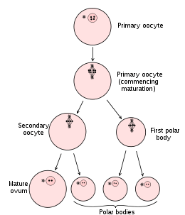
Oogonium —(Oocytogenesis)—> Primary Oocyte —(Meiosis I)—> First Polar Body (Discarded afterward) + Secondary oocyte —(Meiosis II)—> Second Polar Body (Discarded afterward) + Ovum
It should be noted that oocyte meiosis, important to all animal life cycles yet unlike all other instances of animal cell division, occurs completely without the aid of spindle-coordinating centrosomes.[2][3]
The creation of oogonia
The creation of oogonia traditionally doesn't belong to oogenesis proper, but, instead, to the common process of gametogenesis, which, in the female human, begins with the processes of folliculogenesis, oocytogenesis, and ootidogenesis.
Human oogenesis
Oocytogenesis
Oogenesis starts with the process of developing oogonia, which occurs via the transformation of primordial follicles into primary oocytes, a process called oocytogenesis.[4] Oocytogenesis is complete either before or shortly after birth.
Number of primary oocytes
It is commonly believed that, when oocytogenesis is complete, no additional primary oocytes are created, in contrast to the male process of spermatogenesis, where gametocytes are continuously created. In other words, primary oocytes reach their maximum development at ~20[5] weeks of gestational age, when approximately seven million primary oocytes have been created; however, at birth, this number has already been reduced to approximately 1-2 million.
Recently, however, two publications have challenged the belief that a finite number of oocytes are set around the time of birth.[6][7] The renewal of ovarian follicles from germline stem cells (originating from bone marrow and peripheral blood) has been reported in the postnatal mouse ovary. In contrast, DNA clock measurements do not indicate ongoing oogenesis during human females' lifetimes.[8] Thus, further experiments are required to determine the true dynamics of small follicle formation.
Ootidogenesis
The succeeding phase of ootidogenesis occurs when the primary oocyte develops into an ootid. This is achieved by the process of meiosis. In fact, a primary oocyte is, by its biological definition, a cell whose primary function is to divide by the process of meiosis.[9]
However, although this process begins at prenatal age, it stops at prophase I. In late fetal life, all oocytes, still primary oocytes, have halted at this stage of development, called the dictyate. After menarche, these cells then continue to develop, although only a few do so every menstrual cycle.
Meiosis I
Meiosis I of ootidogenesis begins during embryonic development, but halts in the diplotene stage of prophase I until puberty. The mouse oocyte in the dictyate (prolonged diplotene) stage actively repairs DNA damage, whereas DNA repair is not detectable in the pre-dictyate (leptotene, zygotene and pachytene) stages of meiosis.[10] For those primary oocytes that continue to develop in each menstrual cycle, however, synapsis occurs and tetrads form, enabling chromosomal crossover to occur. As a result of meiosis I, the primary oocyte has now developed into the secondary oocyte and the first polar body.
Meiosis II
Immediately after meiosis I, the haploid secondary oocyte initiates meiosis II. However, this process is also halted at the metaphase II stage until fertilization, if such should ever occur. When meiosis II has completed, an ootid and another polar body have now been created.
Folliculogenesis
Synchronously with ootidogenesis, the ovarian follicle surrounding the ootid has developed from a primordial follicle to a preovulatory one.
Maturation into ovum
Both polar bodies disintegrate at the end of Meiosis II, leaving only the ootid, which then eventually undergoes maturation into a mature ovum.
The function of forming polar bodies is to discard the extra haploid sets of chromosomes that have resulted as a consequence of meiosis.
In vitro maturation
In vitro maturation (IVM) is the technique of letting ovarian follicles mature in vitro. It can potentially be performed before an IVF. In such cases, ovarian hyperstimulation isn't essential. Rather, oocytes can mature outside the body prior to IVF. Hence, no (or at least a lower dose of) gonadotropins have to be injected in the body.[11] However, there still isn't enough evidence to prove the effectiveness and security of the technique.[11]
Oogenesis in non-mammals
Some algae and the oomycetes produce eggs in oogonia. In the brown alga Fucus, all four egg cells survive oogenesis, which is an exception to the rule that generally only one product of female meiosis survives to maturity.
In plants, oogenesis occurs inside the female gametophyte via mitosis. In many plants such as bryophytes, ferns, and gymnosperms, egg cells are formed in archegonia. In flowering plants, the female gametophyte has been reduced to an eight-celled embryo sac within the ovule inside the ovary of the flower. Oogenesis occurs within the embryo sac and leads to the formation of a single egg cell per ovule.
In ascaris, the oocyte does not even begin meiosis until the sperm touches it, in contrast to mammals, where meiosis is completed in the estrus cycle.
See also
- Anisogamy
- Archegonium
- Evolution of sexual reproduction
- Female infertility
- Female reproductive system
- Meiosis
- Oncofertility
- Oogonium
- Oocyte
- Origin and function of meiosis
- Sexual reproduction
- Spermatogenesis
References
- ↑ Merriam-Webster Online Dictionary Definition: Oogenesis
- ↑ Szollosi D, Calarco P, Donahue RP (1972). "Absence of centrioles in the first and second meiotic spindles of mouse oocytes". J Cell Sci. 11 (2): 521–541. PMID 5076360.
- ↑ Manandhar G, Schatten H, Sutovsky P (January 2005). "Centrosome reduction during gametogenesis and its significance". Biol. Reprod. 72: 2–13. doi:10.1095/biolreprod.104.031245. PMID 15385423.
- ↑ NCBI - The saga of the germ line
- ↑ Lobo RA (September 2003). "Early ovarian ageing: a hypothesis. What is early ovarian ageing?". Hum. Reprod. 18 (9): 1762–4. doi:10.1093/humrep/deg377. PMID 12923124.
- ↑ Johnson J, Bagley J, Skaznik-Wikiel M, et al. (July 2005). "Oocyte generation in adult mammalian ovaries by putative germ cells in bone marrow and peripheral blood". Cell. 122 (2): 303–15. doi:10.1016/j.cell.2005.06.031. PMID 16051153.
- ↑ Johnson J, Canning J, Kaneko T, Pru J, Tilly J (2004). "Germline stem cells and follicular renewal in the postnatal mammalian ovary". Nature. 428 (6979): 145–50. doi:10.1038/nature02316. PMID 15014492.
- ↑ Forster P, Hohoff C, Dunkelmann B, Schürenkamp M, Pfeiffer H, Neuhuber F, Brinkmann B (2015). "Elevated germline mutation rate in teenage fathers". Proc R Soc B. 282: 20142898. doi:10.1098/rspb.2014.2898. PMC 4345458
 . PMID 25694621.
 - ↑ Biochem
- ↑ Guli CL, Smyth DR (1988). "UV-induced DNA repair is not detectable in pre-dictyate oocytes of the mouse". Mutat Res. 208 (2): 115–119. doi:10.1016/s0165-7992(98)90010-0. PMID 3380109.
- 1 2 Vejledning om kunstig befrugtning 2006 (Danish)
- Bibliography
- Manandhar G, Schatten H and Sutovsky P (2005). Centrosome reduction during gametogenesis and its significance. Biol Reprod, 72(1)2-13.