Trigeminal ganglion
| Trigeminal ganglion | |
|---|---|
 Nerves of the orbit. Seen from above. (Semilunar ganglion visible near bottom.) | |
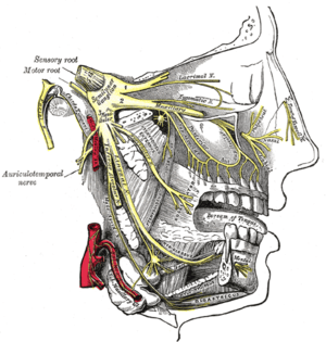 Distribution of the maxillary and mandibular nerves, and the submaxillary ganglion. (Semilunar ganglion visible in upper left.) | |
| Details | |
| Identifiers | |
| Latin | ganglion trigeminale, ganglion semilunare (Gasseri) |
| MeSH | A08.340.390.850 |
| TA | A14.2.01.014 |
| FMA | 52618 |
The trigeminal ganglion (or Gasserian ganglion, or semilunar ganglion, or Gasser's ganglion) is a sensory ganglion of the trigeminal nerve (CN V) that occupies a cavity (Meckel's cave) in the dura mater, covering the trigeminal impression near the apex of the petrous part of the temporal bone.
Structure
It is somewhat crescentic in shape, with its convexity directed forward: Medially, it is in relation with the internal carotid artery and the posterior part of the cavernous sinus.
The motor root runs in front of and medial to the sensory root, and passes beneath the ganglion; it leaves the skull through the foramen ovale, and, immediately below this foramen, joins the mandibular nerve.
The greater superficial petrosal nerve lies also underneath the ganglion.
The ganglion receives, on its medial side, filaments from the carotid plexus of the sympathetic.
It gives off minute branches to the tentorium cerebelli, and to the dura mater in the middle fossa of the cranium.
From its convex border, which is directed forward and lateralward, three large nerves proceed, viz., the ophthalmic (V1), maxillary (V2), and mandibular (V3).
The ophthalmic and maxillary consist exclusively of sensory fibers; the mandibular is joined outside the cranium by the motor root.
Clinical significance
After recovery from a primary herpes infection, the virus is not cleared from the body, but rather lies dormant in a non-replicating state within the trigeminal ganglion.[1]
Herpes Labialis may follow from primary herpes infection/herpetic gingivostomatitis
The trigeminal ganglion is damaged, by infection or surgery, in trigeminal trophic syndrome. Trigeminal trophic syndrome causes paresthesias and anesthesia, which may lead to erosions of the nasal ala.
The thermocoagulation or injection of glycerol into the trigeminal ganglion has been used in the treatment of trigeminal neuralgia.
Other animals
Rodents
In rodents, the trigeminal ganglion is important as it is the first part of the pathway from the whiskers to the brain. Cell bodies of the whisker primary afferents are found here. These afferents are mechanoreceptor cells that fire in response to whisker deflection.
There are around 26,000-43,000 cell bodies in rodent Trigeminal ganglion. It is possible that there are two distinct (or perhaps continuous) populations of cells having slowly and rapidly adapting responses to stimuli.
It is found at the base of the skull and projects to trigeminal brain stem areas including principalis, spinal trigeminal nucleus, interpolaris, and caudalis.
Additional images
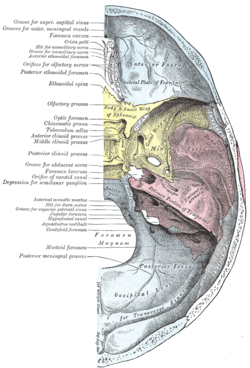 Base of the skull. Upper surface.
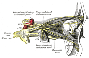 Nerves of the orbit, and the ciliary ganglion. Side view.
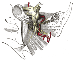 The otic ganglion and its branches.
 Trigeminal ganglion
 Trigeminal ganglion. Deep dissection. Superior view.
References
This article incorporates text in the public domain from the 20th edition of Gray's Anatomy (1918)
- ↑ Verjans GM, Hintzen RQ, van Dun JM, et al. (2007). "Selective retention of herpes simplex virus-specific T cells in latently infected human trigeminal ganglia". Proc. Natl. Acad. Sci. U.S.A. 104 (9): 3496–501. doi:10.1073/pnas.0610847104. PMC 1805572
 . PMID 17360672.
External links
- Diagram at University of Manitoba
- Diagram (as "Gasserian Ganglion") at frca.co.uk
- MedEd at Loyola grossanatomy/dissector/labs/h_n/cranium/cn3_1a.htm
- ancil-484 at NeuroNames
- cranialnerves at The Anatomy Lesson by Wesley Norman (Georgetown University) (V)