Type IV hypersensitivity
| Type IV hypersensitivity | |
|---|---|
 | |
| Video explanation | |
| Classification and external resources | |
| MeSH | D006968 |
Type 4 hypersensitivity is often called delayed type hypersensitivity as the reaction takes two to three days to develop. Unlike the other types, it is not antibody mediated but rather is a type of cell-mediated response.
CD4+ helper T cells recognize antigen in a complex with MHC II major histocompatibility complex on the surface of antigen-presenting cells. These can be macrophages that secrete IL-12, which stimulates the proliferation of further CD4+ Th1 cells. CD4+ T cells secrete IL-2 and interferon gamma, inducing the further release of other Th1 cytokines, thus mediating the immune response. Activated CD8+ T cells destroy target cells on contact, whereas activated macrophages produce hydrolytic enzymes and, on presentation with certain intracellular pathogens, transform into multinucleated giant cells.
Examples
| Disease | Target antigen | Effects |
|---|---|---|
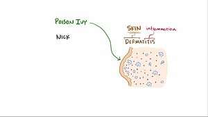
| allergic contact dermatitis[1] | environmental chemicals (e.g., urushiol from poison ivy |
epidermal necrosis, inflammation, skin rash and blisters |
| autoimmune myocarditis[1] | myosin heavy chain protein | cardiomyopathy |
| diabetes mellitus type 1[1] | pancreatic beta cell proteins (possibly insulin, glutamate decarboxylase) |
|
| granulomas[2] | various, depending on underlying disease | walled off lesion containing macrophages and other cells |
| some peripheral neuropathies | Schwann cell antigen | |
| Hashimoto's thyroiditis[1] | thyroglobulin antigen |
hypothyroidism, hard goiter, follicular thymitis |
| inflammatory bowel disease[1] | enteric microbiota and/or self antigens | hyperactivation of T-cells, cytokine release, recruitment of macrophages and other immune cells, inflammation |
| multiple sclerosis[1] | myelin antigens (e.g., myelin basic protein) | myelin destruction, inflammation |
| rheumatoid arthritis[1] | possibly collagen and/or citrullinated self proteins | chronic arthritis, inflammation, destruction of articular cartilage and bone |
| tuberculin reaction | tuberculin |
induration and erythema around injection site indicates previous exposure |
An example of a TB infection that came under control: M. tuberculosis are engulfed by macrophages after being identified as foreign, but due to an immuno-escape mechanism peculiar to mycobacteria, TB bacteria are able to block the fusion of their enclosing phagosome with lysosomes which would destroy the bacteria. Thereby TB can continue to replicate within macrophages. After several weeks, the immune system somehow [mechanism as yet unexplained] ramps up and, on stimulation with IFN-gamma, the macrophages become capable of killing M. tuberculosis by forming phagolysosomes and nitric oxide radicals. However the hyper-activated macrophages secrete TNF which recruits multiple monocytes into the battle. These cells differentiate into epithelioid histiocytes which wall off the infected cells, but at the cost of significant inflammation and local damage.
Some other clinical examples:
- Temporal arteritis
- Symptoms of leprosy
- Symptoms of tuberculosis
- Coeliac disease
- Graft-versus-host disease[4]
- Chronic transplant rejection
See also
References
- 1 2 3 4 5 6 7 Kumar, Vinay; Abbas, Abul K.; Aster, Jon C. (2012-05-01). Robbins Basic Pathology. Elsevier Health Sciences. ISBN 1455737879.
- ↑ "Hypersensitivity reactions". www.microbiologybook.org. University of South Carolina School of Medicine - Microbiology and Immunology On-line. Retrieved 2016-05-29.
- ↑ "Hypersensitivity reactions". www.microbiologybook.org. Retrieved 2016-05-29.
- ↑ "eMedicine - Hypersensitivity Reactions, Delayed : Article by Walter Duane Hinshaw".