Anterior chamber of eyeball
| Anterior chamber of eyeball | |
|---|---|
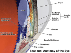
 Anterior part of human eye, with anterior chamber at right. | |
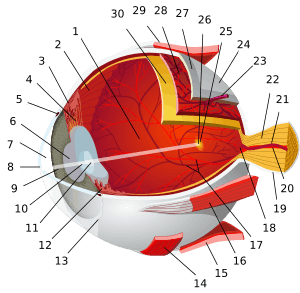
 Schematic diagram of the human eye. | |
| Details | |
| Identifiers | |
| Latin | camera anterior bulbi oculi |
| TA | A15.2.06.003 |
| FMA | 58078 |
The anterior chamber (AC) is the fluid-filled space inside the eye between the iris and the cornea's innermost surface, the endothelium.[1] Aqueous humor is the fluid that fills the anterior chamber. Hyphema and glaucoma are two main pathologies in this area. In hyphema, blood fills the anterior chamber. In glaucoma, blockage of the canal of Schlemm prevents the normal outflow of aqueous humor, resulting in accumulation of fluid, increased intraocular pressure, and eventually blindness. The normal depth of anterior chamber of eye is 3.5mm to 2.5mm; less than 2.5mm depth can be risk for angle closure glaucoma.
One peculiar feature of the anterior chamber is dampened immune response to allogenic grafts. This is called anterior chamber associated immune deviation (ACAID), a term introduced in 1981 by Streilein et al.[2][3]
Pathology
Additional Images
-

Structures of the eye labeled
-

This image shows another labeled view of the structures of the eye
See also
References
- ↑ Cassin, B.; Solomon, S. (1990). Dictionary of eye terminology. Gainesville, Fla: Triad Pub. Co. ISBN 0-937404-33-0.
- ↑ Streilein JW, Niederkorn JY (May 1981). "Induction of anterior chamber-associated immune deviation requires an intact, functional spleen". J. Exp. Med. 153 (5): 1058–67. doi:10.1084/jem.153.5.1058. PMC 2186172
 . PMID 6788883.
 - ↑ "Archived copy". Archived from the original on 2015-02-11. Retrieved 2012-07-16.
External links
- Atlas image: eye_2 at the University of Michigan Health System - "Sagittal Section Through the Eyeball"

